Method for discriminating red blood cells from white blood cells by using forward scattering from a laser in an automated hematology analyzer

Inactive Publication Date: 2010-10-28
ABBOTT LAB INC
View PDF44 Cites 70 Cited by
  • Summary
  • Abstract
  • Description
  • Claims
  • Application Information

AI Technical Summary

Benefits of technology

[0017]The use of a laser having a wavelength ranging from about 400 nm to about 450 nm aids in the separation of red blood cells from white blood cells. The use of the aforementioned laser enables the hematology analyzer to function without the need for a lysing agent for red blood cells in order obtain an accurate count of white blood cells.
[0018]In one embodiment, the analyzer uses one single dilution of whole blood to count all of the cells in the sample. This count is achieved by using two different sample introduction rates for the diluted blood into the flow cell. The white blood cell count uses a sufficiently high sample introduction rate and an intermediate angle scattering (IAS) trigger in order to exclude platelets and red blood cells. The overall sample introduction rate for counting red blood cells and platelets is lower than the overall sample introduction rate used for counting white blood cells. This technique allows a sufficient number of white blood cell events to be collected in less than 40 seconds of counting time. Introduction of the diluted sample into the flow cell can be carried out by injection. The actual rates of sample introduction can be determined by one of ordinary skill in the art without undue experimentation. For example, in certain embodiments a low rate of sample introduction may be in the range of about 1,000-50,000 cells per second (for samples with typical concentrations found in normal adult blood); while in certain embodiments a high rate of sample introduction may be in the range of about 50,001-300,000 cells per second (for samples with typical concentrations found in normal adult blood).

Problems solved by technology

However, the use of lysing agents to lyse red blood cells brings about certain difficulties and complications in the detection of white blood cells.
The lysing agent may be insufficiently strong, thereby resulting in red blood cells being counted as white blood cells.
Alternatively, the lysing agent may be excessively strong, thereby resulting in artificially low counts of white blood cells.
In hematological assays aimed at determining parameters from human whole blood, there are two physiological factors that present obstacles to simple, rapid, and accurate determination of cell counts.
Therefore, when determining red blood cells mainly by size discrimination, the asymmetry in concentration can work in one's favor, since the occasional white blood cell misclassified as a red blood cell will not, generally, affect the overall accuracy of the measured concentration of red blood cells to any appreciable degree; however, the converse is not true, and any unaccounted-for interference from red blood cells in determining the concentration of lymphocytes (and, by extension, the overall concentration of white blood cells) can yield very inaccurate results.
Both of these methods have drawbacks.
First of all, the lysing reagent used to dissolve the red blood cells can attack the white blood cells as well, reducing their integrity and eventually dissolving them, too.
This is particularly a problem with white blood cells that are already fragile in the first place, due to some pathological condition (such, as, for example, chronic lymphocytic leukemia).
This tailoring, however, is a complex solution, which additionally either requires prior knowledge of the state of the patient, or must be used as a reflex test following a standard complete blood count (CBC).
Regarding the fluorescence-based approach at discriminating between red blood cells and lymphocytes, the main obstacle is the measurement rate.
This throughput is far lower than that achievable by most automated hematology analyzers commercially available today, severely limiting its commercial usefulness.
While it would technically be possible to count the white blood cells using this same assay, as they are sufficiently differentiated by fluorescence from either red blood cells or reticulocytes to obtain the desired accuracy, the relatively low concentration of white blood cells in the dilution used makes it an impractical option to achieve the required statistical precision.
All of these benefits, however, are overshadowed in the prior art by the low throughput of the disclosed method.

Method used


Image

  • Method for discriminating red blood cells from white blood cells by using forward scattering from a laser in an automated hematology analyzer
  • Method for discriminating red blood cells from white blood cells by using forward scattering from a laser in an automated hematology analyzer
  • Method for discriminating red blood cells from white blood cells by using forward scattering from a laser in an automated hematology analyzer

Examples


example 1

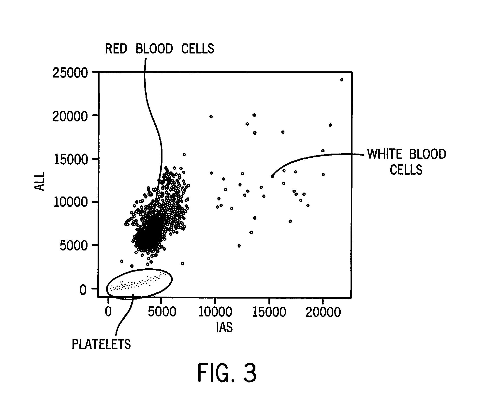
[0102]This example illustrates separation of red blood cells, white blood cells, and platelets in a sample of unlysed whole blood carried out on a prototype analyzer that uses a laser having a wavelength of 405 nm. The results are shown in FIG. 3. It can be seen that there is clear separation between platelets, red blood cells, and white blood cells. The number of white blood cells is too low to see a white blood cell differential. In order to collect more white blood cell events, an IAS trigger value corresponding to about 8000 on the IAS is required.

example 2

[0103]This example illustrates a white blood cell differential carried out on a prototype analyzer that uses a laser having a wavelength of 405 nm, wherein the method does not employ a lysing agent. The results are shown in FIG. 4, where monocytes are designated by the letter “M”, lymphocytes are designated by the letter “L”, neutrophils are designated by the letter “N”, and eosinophils are designated by the letter “E.” The classification is achieved without a clustering algorithm.

example 3

[0104]This example illustrates that issues encountered with lysis-resistant red blood cells interfering with the white blood cell count by the CELL-DYN® Ruby® hematology analyzer and the CELL-DYN® Sapphire® hematology analyzer are significantly improved with a laser having a wavelength of 405 nm. CELL-DYN® Ruby® hematology analyzers employ a wavelength of 633 nm. CELL-DYN® Sapphire® hematology analyzers employ a wavelength of 488 nm. The same blood sample was run on three different analyzers using four separate assays; the results are described below, with reference to FIGS. 5-8.

[0105]FIGS. 5A, 5B, 5C, and 5D show that the results generated by the analyzer would indicate that the sample has a lymphocyte percentage of 95%, as measured with a CELL-DYN® Sapphire° hematology analyzer employing a laser having a wavelength outside the range of 400 nm to 450 nm and a lysing agent of low lytic strength. FIGS. 6A, 6B, 6C, and 6D show that the results generated by the analyzer would indicate ...


PUM

PropertyMeasurementUnit
Angleaaaaaaaaaa
Angleaaaaaaaaaa
Angleaaaaaaaaaa

Abstract

A method for identifying, analyzing, and quantifying the cellular components of whole blood by means of an automated hematology analyzer and the detection of the light scattered, absorbed, and fluorescently emitted by each cell. More particularly, the aforementioned method involves identifying, analyzing, and quantifying the cellular components of whole blood by means of a light source having a wavelength ranging from about 400 nm to about 450 nm and multiple in-flow optical measurements and staining without the need for lysing red blood cells.

Description

BACKGROUND OF THE INVENTION[0001]1. Field of the Invention[0002]Certain embodiments of this invention relate to a method for identifying, analyzing, and quantifying the cellular components of whole blood by means of an automated hematology analyzer and the detection of the light scattered and absorbed by each cell. More particularly, the aforementioned method may involve identifying, analyzing, and quantifying the cellular components of whole blood by means of multiple in-flow optical measurements using a single dilution of sample without the need for lysing red blood cells.[0003]2. Discussion of the Art[0004]Some automated hematology analyzers are equipped with an optical bench that can measure multiple in-flow optical measurements, such as light scattering, extinction, and fluorescence, as described in U.S. Pat. Nos. 5,631,165 and 5,939,326, both of which are incorporated herein by reference. Furthermore, U.S. Pat. Nos. 5,516,695 and 5,648,225, both of which are incorporated herei...

Claims


Application Information

Patent Timeline
IPC IPC(8): C12Q1/68C12M1/34C12Q1/06
CPCG01N15/00G01N33/49G01N2015/0073G01N2015/0076Y10T436/101666G01N2015/0084G01N2015/1486G01N15/1459G01N2015/008G01N33/4915G01N33/492G01N2015/014G01N2015/018G01N2015/012G01N2015/016G01N21/53G01N15/1404G01N15/1429G01N2015/1006G01N2800/22
Inventor KROCKENBERGER, MARTINWU, JIONGROEMER, BODOVACCA, GIACOMO
Owner ABBOTT LAB INC
Why Eureka
Try Eureka