
Method of contactless magnetic electroporation

a magnetic electroporation and contactless technology, applied in the field of contactless magnetic electroporation, can solve the problems of insufficient quantity or size of pores, apoptosis or cell lysis, pain, infection risk, etc., and achieve the effect of not having an effect on the efficiency of transfection

Inactive Publication Date: 2010-09-30
INOVIO PHARMA
View PDF7 Cites 29 Cited by
  • Summary
  • Abstract
  • Description
  • Claims
  • Application Information

AI Technical Summary

Benefits of technology

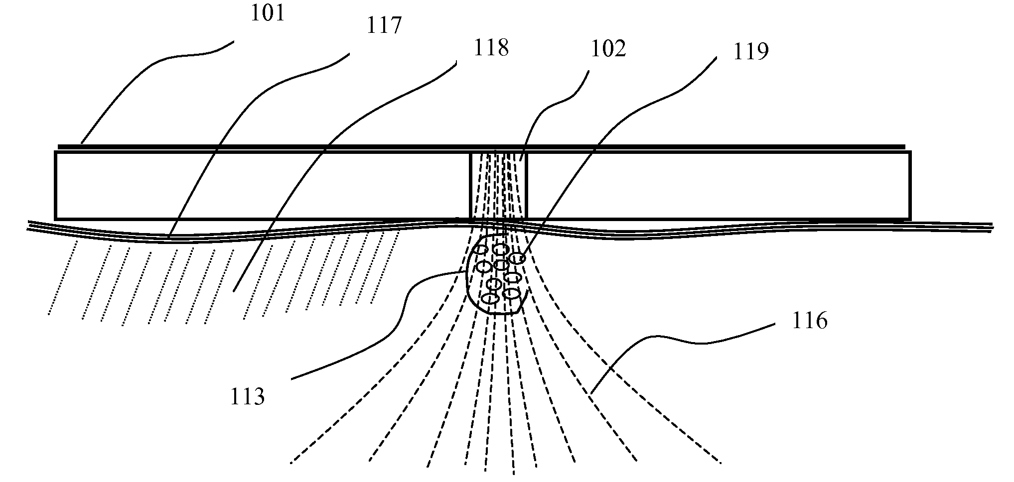
The patent describes a new method for inducing membrane poration without using traditional electrodes. Instead, a changing magnetic field is used to directly induce electrical and ionic currents, which then facilitate the formation of membrane pores. The magnetic field acts as a substitute for an electric field, and the induced currents are measured in units of amps per unit of area. The method is not dependent on the resistance of the tissue, which makes it more efficient than traditional electroporation. The patent also describes the use of magnetic flux density to measure the effect of the magnetic field on membrane poration. The invention has potential applications in biological tissue and can be used to transfer agents into cells without the need for traditional electrodes.

Problems solved by technology

Pulses of high field strength and long duration generally will induce a large quantity and / or pores of large size that may lead to apoptosis or cell lysis.
Conversely, pulses of low field strength and / or short duration may induce an insufficient quantity or size of pores that will allow only a low flux rate of ions and molecules across the cell membrane.
Penetrating electrodes tend to use sharp needles that break through the stratum corneum barrier and into or through the dermis, which causes trauma, pain and risk of infection.
Such tissue-piercing needle electrodes also require a complex manufacturing process governed by medical device manufacturing regulations and further involve sterilization and the need to maintain sterile packaging, thereby causing a significant cost per patient use.
This technique has been successful in delivering macromolecules past cell membranes into the cytoplasm, but also requires a complex manufacturing process to successfully apply the macromolecules onto the gold beads or other particles in sufficient density.
Moreover, the consistency of skin can vary greatly from individual to individual and the amount of agent that can be deposited on the carrier particles is relatively small, presenting a major challenge for consistent and sufficient delivery of an agent.
Attachment of a magnetic bead made of a ferrous core or other magnetically active metal to an agent molecule is a complicated process, and delivery of such a particle-agent complex may leave a composite molecule within cells and tissues that may cause unwanted side effects.
Additionally, U.S. Pat. No. 6,132,419 discusses the possibility of using an inductance device for introduction of molecules into living cells; however, it does not provide any parameters, data or reduction to practice.
However, as compared to muscle twitches generated by electroporation with traditional penetrating needles, the muscle movement is uninhibited and does not endanger the twitching tissue.
Movement of skin or muscle tissue while it contains embedded sharps causes tissue tearing and may result in additional trauma and pain over and above the initial insertion of the sharps.
Compared to the understanding and applications of electric fields in living systems, there is a relative void in the understanding and uses of magnetic fields in living systems.

Method used


Image

  • Method of contactless magnetic electroporation
  • Method of contactless magnetic electroporation
  • Method of contactless magnetic electroporation

Examples


example 1

[0054]Experimental evidence of effective magnetopermeabilization was demonstrated by 5 sec series of monophasic pulses delivered at the rate of 10 pulses / sec for a total of 50 pulses per DNA injection site. Six injection sites were prepared, each with an intradermal injection of approximately 20 microliters of 1 mg / ml concentration gWiz-GFP (Green Fluorescent Protein plasmid from Aldevron LLC, Fargo, N. Dak.). The rise time of each magnetic pulse from zero to approximately 4 tesla was achieved within 1 microsecond, for a magnetic field rate-of-change of at least 4 tesla / microsecond. This actual pulse pattern is shown in FIG. 13b. The experimental results in terms of plasmid DNA expression for monophasic pulses are seen on the left side of FIG. 15c.

example 2

[0055]Similar experimental evidence of magnetopermeabilization was demonstrated by 5 sec series of biphasic pulses delivered at the rate of 10 pulses / sec, for a total of 50 pulses per DNA injection site. Six injection sites were prepared, each with an intradermal injection of approximately 20 microliters of 1 mg / ml concentration gWiz-GFP (Green Fluorescent Protein plasmid from Aldevron LLC, Fargo, N. Dak.). The rise time of each magnetic pulse from zero to approximately 4 tesla was within 1 microsecond, for a magnetic field rate-of-change of at least 4 tesla / microsecond. This actual pulse pattern is shown in FIG. 14c. An expanded time base in FIG. 14d indicates the rise time closer to 400 nanoseconds, which equates to a magnetic field rate-of-change to 4 tesla / 400 ns=10 tesla / microsecond. The experimental results in terms of DNA expression for biphasic pulses are seen on the right side of FIG. 15c. When fewer pulses were delivered per DNA injection site, such as comparing 20 pulses ...


PUM


Abstract

This invention provides a novel method of tissue electroporation that eliminates the need for electrodes that conduct electricity to the tissues. This invention creates electric currents and fields sufficient for porating cell membranes for improving the delivery of polynucleotides such as plasmid and linear DNA and RNA constructs, and polypeptides such as antigen protein constructs into mammalian eucaryotic cells purely by magnetic field pulses that does not require the use of contacting electrodes to conduct electric or ionic current. This invention thus provides a method for improving transfection and immunogenicity of pharmaceutical substances without direct contact with a living body, and may be called magnetopermeabilization. A concomitant aspect of the invention is the method by which a drug such as a solution containing DNA is delivered to a targeted tissue bed that is optimal in conjunction with magnetopermeabilization for maximal transgene expression and drug effect.

Description

[0001]This application claims priority to Provisional Application 61 / 164,471 filed Mar. 30, 2009.[0002]Assignment: MagneGene, Inc., a California Corporation.FIELD OF THE INVENTION[0003]The present invention relates to the method of delivery of therapeutic substances including polynucleotides and polypeptides into mammalian eukaryotic cells by inducing permeabilization of cell membranes with magnetic pulses. Particularly, the present invention relates to permeabilizing tissues near the surface of the body such as dermis, epidermis, sub-dermal regions, muscle tissues and tumor tissues by magnetically inducing electroporation without placement of electrodes onto, or within these tissues that conduct electricity to said tissues. Therefore, the present invention provides for a novel method for electroporation of cells that does not require physical contact with said tissues and thus does not require sterile or non-sterile electrodes to deliver energy. This method is related to the princi...

Claims


Application Information

Patent Timeline
Patent Type & Authority Applications(United States)
IPC IPC(8): A61N2/04A61N2/00
CPCA61N2/02A61N2/002
Inventor KARDOS, THOMAS JOSEPHRABUSSAY, DIETMAR PAUL
Owner INOVIO PHARMA
Learn More