Laser scanning digital camera with pupil periphery illumination and potential for multiply scattered light imaging
- Summary
- Abstract
- Description
- Claims
- Application Information
AI Technical Summary
Benefits of technology
Problems solved by technology
Method used
Image

Examples
Embodiment Construction
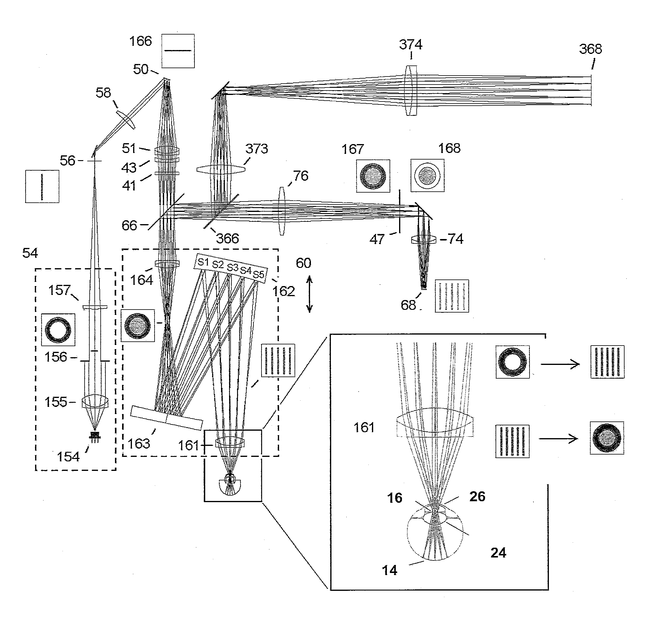
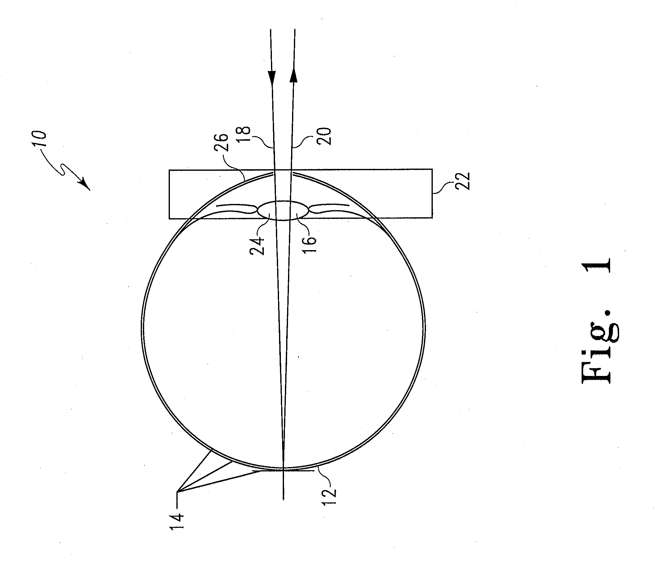
[0055]The present application relates to a small, portable lightweight instrument or device of low cost particularly suitable for examining the retinal and subretinal layers of the eye 10 (see FIG. 1) for abnormalities. The device is noncontact and does not require drops to dilate the pupil of the eye in order to obtain a retinal or sub-retinal image. Referring to FIG. 1, the plane of focus 12 of the device includes the vitreoretinal interface and the retinal nerve fiber layer 13, which have the greatest amount of light return of the several sublayers of the retina 14. This area of greatest light return is helpful in finding the plane of focus, but presents a problem when trying to image through that area of the retina 14. When a human retina 14 is imaged, light from an illumination source is passed through a slit aperture to produce a line source and is scanned across a desired focal plane in the eye after passing through the entrance pupil 16. Light enters through one or more port...
PUM
 Abstract
Description
Claims
Application Information
 - R&D
- Intellectual Property
- Life Sciences
- Materials
- Tech Scout
- Unparalleled Data Quality
- Higher Quality Content
- 60% Fewer Hallucinations
Browse by: Latest US Patents, China's latest patents, Technical Efficacy Thesaurus, Application Domain, Technology Topic, Popular Technical Reports.
© 2025 PatSnap. All rights reserved.Legal|Privacy policy|Modern Slavery Act Transparency Statement|Sitemap|About US| Contact US: help@patsnap.com