
X-Ray Machine for Breast Examination Having a Beam Configuration for High Resolution Images

a breast and beam configuration technology, applied in the field of x-ray machines for breast examination, can solve the problem that the relative large breast wall cannot be imaged with x-ray, and achieve the effect of high resolution and favorable cos

Active Publication Date: 2010-04-01
AB CT ADVANCED BREAST CT GMBH
View PDF72 Cites 10 Cited by
  • Summary
  • Abstract
  • Description
  • Claims
  • Application Information

AI Technical Summary

Benefits of technology

[0007]An objective of the disclosure provided herein is to design an X-ray machine, which images a female breast in a diagnostically correct manner, rapidly, at favorable cost, and with high resolution. Another object of the disclosure is to design an X-ray detector for an X-ray machine of this kind.

Problems solved by technology

However, a relatively large region of a wall of a breast cannot be imaged with the X-ray machine disclosed in the US publication.

Method used


Image

  • X-Ray Machine for Breast Examination Having a Beam Configuration for High Resolution Images
  • X-Ray Machine for Breast Examination Having a Beam Configuration for High Resolution Images
  • X-Ray Machine for Breast Examination Having a Beam Configuration for High Resolution Images

Examples


Embodiment Construction

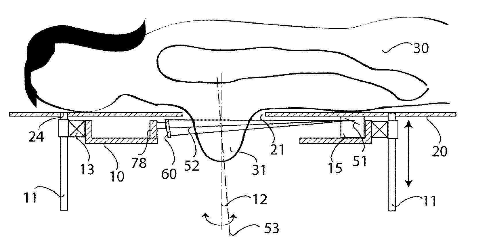
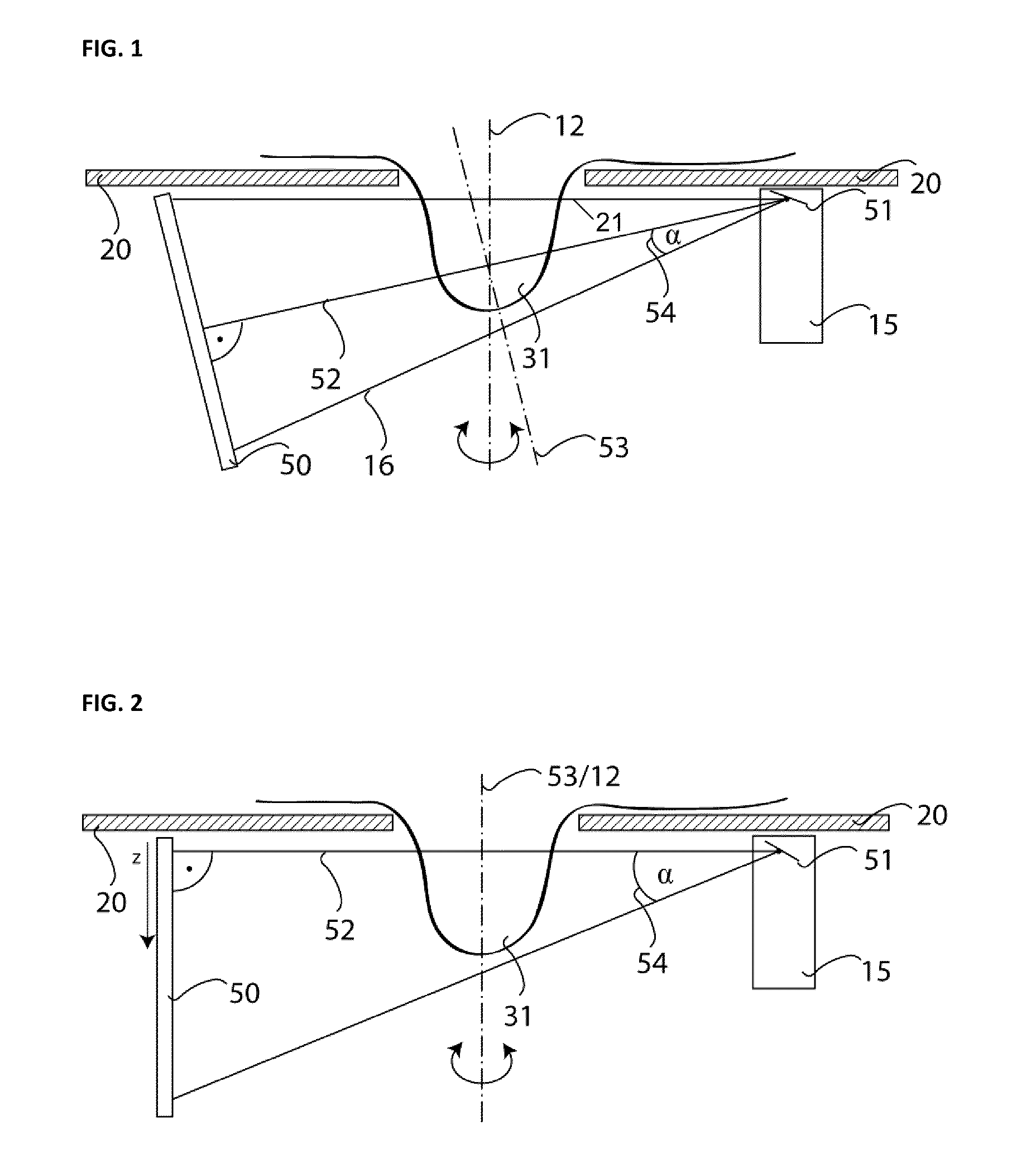
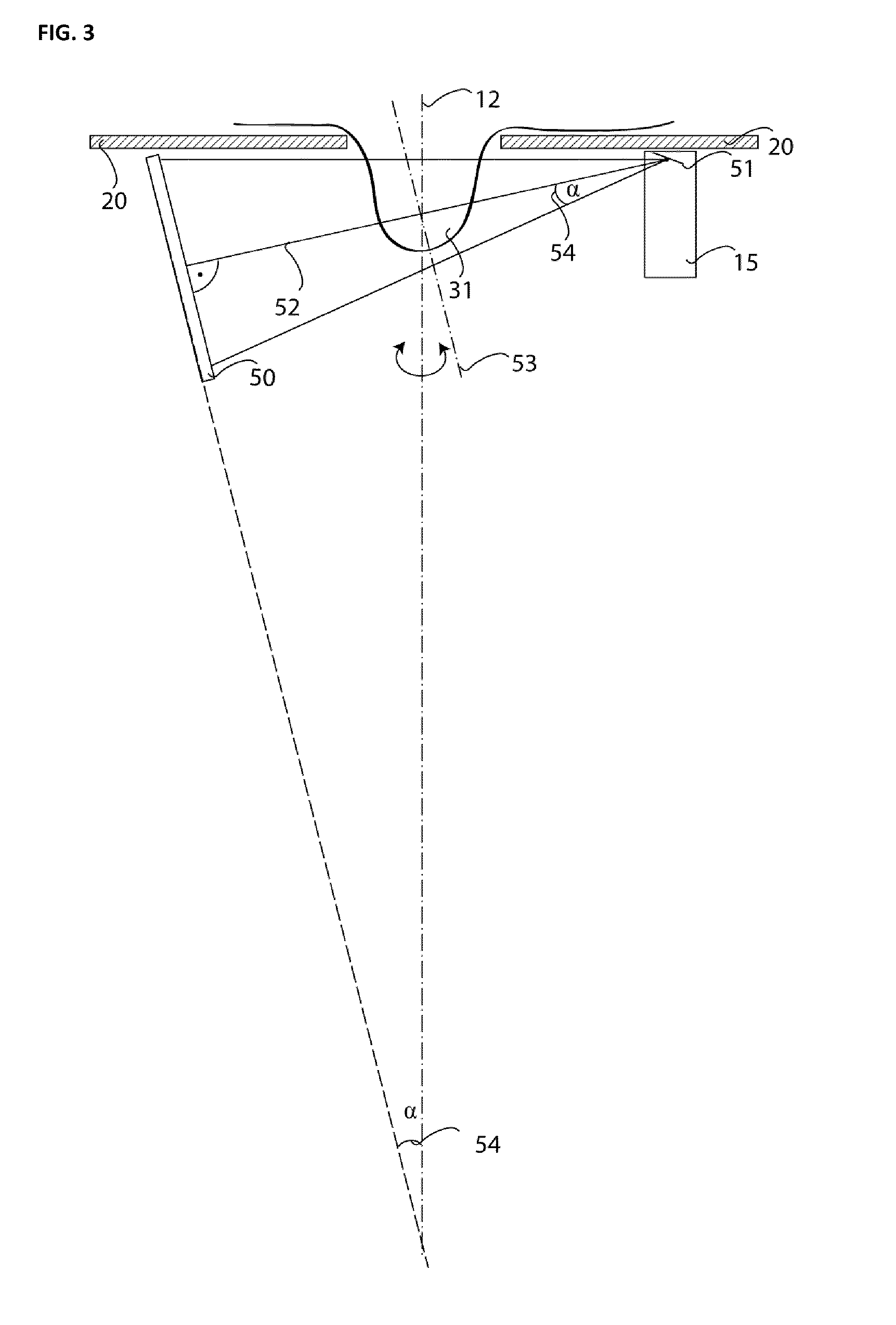
[0025]FIG. 1 illustrates an embodiment of an X-ray machine having a flat panel detector 50. A patient's breast 31 to be examined is suspended through an opening 21 in a support surface, which in this embodiment is a patient table 20. Of course, this arrangement may also be rotated through a range of desired angles, resulting in an alternative arrangement in which the patient table 20 is inclined or stood up on end. In such an arrangement, the inclined or stood up patient table may only be an abutting surface through which a breast is inserted. In either embodiment, it is desired that chest wall of the patient rest as close as possible to the patient table 20, so that the breast can be imaged as completely as possible.

[0026]The X-ray machine shown in FIG. 1 includes a gantry (not shown in FIG. 1) rotating about a rotational axis 12. Among other components, the rotating gantry includes an X-ray tube 15 and a flat panel detector 50. The anode 51 within the X-ray tube 15 generates a bea...


PUM


Abstract

An X-ray machine for imaging a breast of a female patient includes a patient table for accommodating the patient, an X-ray tube and an X-ray detector. In one embodiment, the X-ray machine may be designed as a spiral CT scanner having a rotatable gantry on which the X-ray tube and detector are mounted. The X-ray detector is inclined with respect to the X-ray tube, so that a central ray of a beam of rays emitted from the X-ray tube is perpendicularly incident on an active face of the detector. In one embodiment, the detector may be designed with a curvature, which reduces image artifacts by reducing path length differences between the central ray and the outer rays of the beam. This configuration provides particularly space saving X-ray machines, which at the same time, are of particularly high resolution.

Description

PRIORITY CLAIM[0001]This application claims priority to pending German Application No. DE102008042430.7 filed on Sep. 29, 2008.BACKGROUND OF THE INVENTION[0002]1. Field of the Invention[0003]The invention relates to an X-ray machine for imaging a female breast (mammography). In particular, the invention relates to a machine with which high-resolution X-ray exposures may be produced.[0004]2. Description of Related Art[0005]X-ray machines with an X-ray tube and a detector of large surface are known from prior art. For example, an X-ray machine is disclosed in U.S. Pat. No. 6,480,565, in which the size of the detector is dimensioned so that an entire breast can be imaged. In order to produce three-dimensional images, the X-ray tube and the detector are located on a rotatable gantry, which permits exposures to be made from different angular positions. Another X-ray machine, which is based on a spiral computer tomography (CT) scanner, is disclosed in U.S. Publication No. 2007 / 0064867. In...

Claims


Application Information

Patent Timeline
Patent Type & Authority Applications(United States)
IPC IPC(8): A61B6/04
CPCA61B5/015A61B6/027A61B5/704A61B6/032A61B6/035A61B6/0435A61B6/06A61B6/107A61B6/502A61B6/583A61B18/00A61B2017/00084A61B2019/205A61B2019/5238A61B2019/524G01K11/30G06T7/0012G06T7/0083G06T2207/10081G06T2207/30068G06T2207/30096A61B5/4312A61B6/4275A61B90/17A61B2090/376A61B2090/3762G06T7/74G06T7/12
Inventor KALENDER, WILLISCHILLING, HARRY
Owner AB CT ADVANCED BREAST CT GMBH
Learn More