
Method for the treatment of skin tissues

a skin tissue and tissue technology, applied in the field of skin tissue treatment, can solve the problems of difficult to deliver relatively large molecules using this approach, many compounds do not penetrate the skin at all, and difficult route of drug administration, so as to reduce irregular cold and hot spots, effective careful activation, and thermal peak effect of the photosensitizer

Inactive Publication Date: 2009-10-01
BAROLET DANIEL
View PDF2 Cites 27 Cited by
  • Summary
  • Abstract
  • Description
  • Claims
  • Application Information

AI Technical Summary

Benefits of technology

[0033]IR exposure as described herein is a new way to deliver, for example through a substantially uniform penetration, a given compound in the skin. The opening of pores takes place without mechanical manipulation or alteration of skin integrity. The nature of the substance or compound might have less influence over this delivery procedure as this invention refers to the induction of a physiological process: heat generated pore dilatation and physicochemical permeation (to pass through epidermal openings or interstices) secondary to photobiochemical vibrational alterations. As oppose to existing mechanical (microdermabrasion) or purely chemical (acetone scrub) pre-PDT skin preparations, the present method uses non-ablative IR photons non-invasively to achieve inside-out thermal benefits without any damage to skin. A further benefit of this invention over existing technologies is the development of a well controlled, users friendly, and consistent procedure, easy to use in a clinical setting.
[0034]The radiant IR skin preparation method provides numerous advantages. The inside-out heat transfer mechanism proper to this method does not imply skin contact with a light source, providing uniformity in the preparation of the entire treatment area. Moreover, this method, while triggering a physiological reaction at the treatment site, is independent of the molecule size and drug rate for transcutaneous delivery. And as this skin preparation method occurs prior to any drug application, it cannot alter the drug integrity in any mean. Finally, IR radiant skin preparation can be performed from up to an hour before treatment in order to open skin pore for optimal drug delivery, according to selected irradiation parameters.
[0035]This innovative method provides of relatively quick and easy way to enhance drug absorption as IR exposure tries to reproduce the human body normal physiological reactions for heat dissipation, leading to the opening of skin pores.
[0037]First, the use of an LED source avoids, or at least reduces, thermal peak effect on the photosensitizer—so called thermal effects—usually encountered with thermal technologies such as IPLs and lasers (i.e. PDL; Pulsed Dye Lasers). LED technology clearly allows for progressive photoactivation of photosensitizers. Furthermore, dose-rate is increasingly believed to be one of the important criteria as opposed to total dose (fluence). Uniformity must also be addressed as a high power LED light source covering large treatment areas must reduce irregular cold and hot spots. A high power non-thermal device offers the threshold energy level required for effective careful activation of the photosensitizer with minimal side effects. In addition, the wavelength specification is key to matching selective absorption peaks of the photosensitizer—a wavelength with a narrow spectral band reaching deeper dermal structures should be used in many instances. In fact, the use of a dual wavelength (red and blue) LED light source enhances PDT results for acne and other sebaceous disorders. Red wavelength (630 nm) can reach the sebaceous glands and blue (405 nm) photobleaches any residual protoporphyrin IX (PpIX) in the epidermis, thereby also reducing post-treatment photosensitivity. Indeed, a dual-wavelength LED device optimizes PDT results by providing a superior activation of the photosensitizer—deep at the target structure—for maximized clinical effect and fewer side effects.

Problems solved by technology

Many compounds will be absorbed by the skin; however the absorption typically involves relatively small quantities / concentrations of external molecules per area of skin, per hour, requiring that unpractical large skin contact areas be used to achieve therapeutically effective concentrations of substances via transcutaneous delivery.
Furthermore, many compounds do not penetrate the skin at all.
Transcutaneous delivery remains to these days a challenging route of drug administration.
A typical challenge faced with inhalable or oral delivery is drug concentration, as it regards the delivery of sufficient quantities of the drug to relatively inaccessible inner surfaces (internal organs) where the delivered compound crosses into the blood.
However, this method requires that the molecules used be charged, which is not automatically the case for all substances of interest.
It is also relatively difficult to deliver relatively large molecules using this approach.
Although this method may be useful to allow some drug molecules to reach dermal capillaries, there is no evidence that it would promote preferential absorption and deposition to specific target structures within the dermis.
The main limitation of this technology is the depth of penetration of these channels within the epidermis so that not enough drug molecules are able to get to targeted structures in the dermis to achieve a significant clinical improvement.
However, the PDT treatment disclosed in this document is invasive in nature and no transcutaneaous delivery of the photosensitizer is therefore contemplated as the radiation is applied through a probe inserted within the tissues to be treated.
The need for repeated microtrauma to the skin, the requirement of sometimes large contact areas to achieve proper drug concentration and the need for a patch with prolonged contact time are all disadvantages of this method.
Sound waves create cavitations bubbles in the tissue, disrupting the lipid bilayers of stratum corneum cells, which results in the creation of microchannels.
The limitations of this method are the same as for the ones described previously for heat.

Method used


Image

  • Method for the treatment of skin tissues
  • Method for the treatment of skin tissues
  • Method for the treatment of skin tissues

Examples


example 1

Radiant IR Increases Skin Temperature and Opens Skin Pores (Anterior Arm)

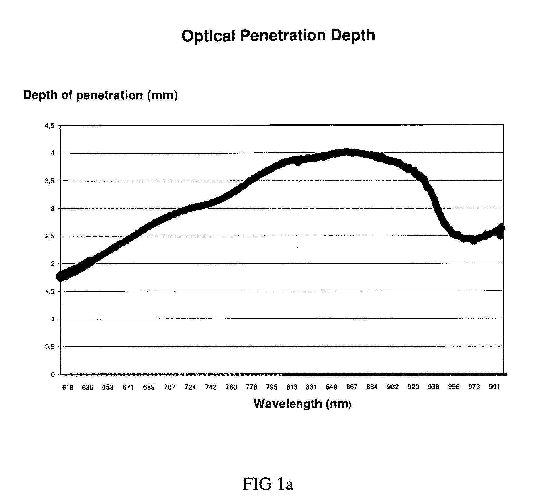
[0063]A thermocouple type-T probe (Omega inc.) was inserted at the papillary junction of the skin (D-E (dermo-epidermal) junction) of the anterior arm of a subject to allow real time measurements of skin temperature during IR exposure. Preliminary testing performed on an ex vivo animal model had shown a significant increase in temperature using radiation of 870 nm, at 80 mW / cm2, with a source 3 cm away from the target area for exposures up to 30 minutes (resulting in a fluence of up to 144 J / cm2) (data not shown). The human model (in vivo) testing described herein considers superior tissue mass (bulk effect) and inherent physiological body temperature management mechanisms (i.e. blood capillaries heat dissipation) that could influence the temperature variation monitored by the probe during IR exposure.

[0064]Two sets of parameters were investigated: 870 nm at 200 mW / cm2 (High Irradiance) and 970 nm at 50 mW / cm2 ...

example 2

Radiant IR Skin Preparation: Temperature and Sebum Monitoring

[0065]The treatment area, the middle upper back, was cleaned with a mild soap 60 min prior to the experiment. Sebum measurement was assessed with a dermaspectrometer and temperature was monitored with a thermocouple (Omega inc.). Then, 15 min of 870 nm IR light, delivered 117 J, at a 2.5 cm treatment distance (130 mW / cm2, Mode: continuous wave (CW).). Sebum readings using a sebumeter SM815 (CK electronic GmbH) were taken prior to irradiation (T0), after 15 min irradiation time (T15), right after the acetone scrub (Ta), after the second 15 min irradiation time (T215), and at 30 min cool off time (Tc30). The following readings were measured on the upper back: T0:8; T15:13; Tc30:11. Periodic epidermal temperature was monitored every minute during IR exposure using a type T thermocouple firmly resting on the epidermis, in the middle of the treatment area on the upper back. Temperature was monitored every minute. During this ex...

example 3

IR Skin Preparation Leads to Temperature Increase (Middle Upper Back) Enhancing PDT Treatment Outcome in an Acne Patient

[0066]A treatment was carried on a 32-year-old female suffering from mild inflammatory acne in the upper back. Briefly, the treatment area was cleaned with a mild soap and an acetone scrub was performed. The IR-device (870 nm, 130 mW / cm2, Mode: CW) was placed over the treatment area (middle upper back). A distance gauge maintained the treatment distance at 2.5 cm during the entire procedure. Then, the treatment area was exposed to the IR LED device for 15 min and 177 J were delivered. After the radiant IR skin preparation, the treatment area and the photosensitizer, kept at room temperature, was applied for 90 min. Then, 5-aminolevulinic acid (Levulan™ Kerastick™) was activated by red LED light delivered by the LumiPhase-R (Opusmed, Canada), for 20 min. Finally, a 5 min exposure at 405 nm LED 30 mW / cm2) was performed. Her back was then washed-off and the patient wa...


PUM


Abstract

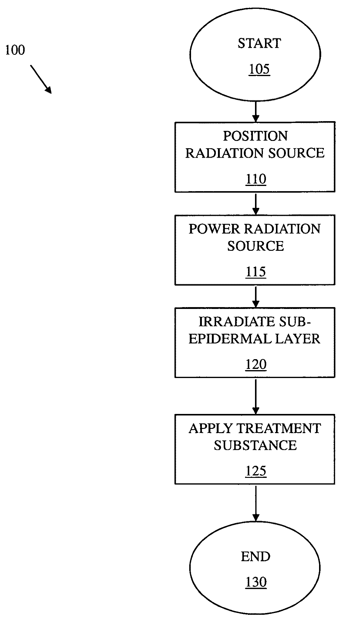
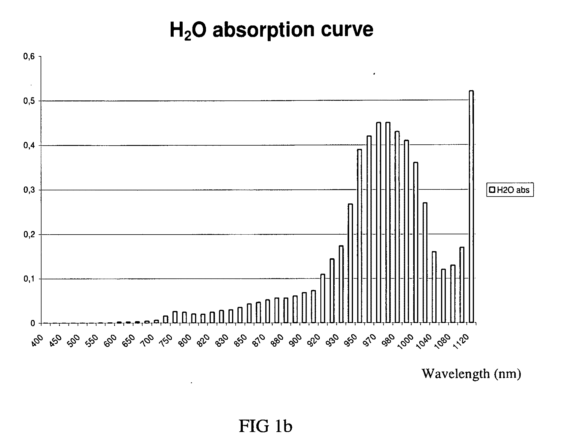
A method for treating skin tissues, the skin tissues defining an epidermal layer and a sub-epidermal layer, the epidermal layer defining a skin surface and the sub-epidermal layer extending from the epidermal layer substantially opposite to the skin surface, the method comprising: positioning a radiation source outside of the skin tissues at a predetermined distance from the skin surface; powering the radiation source so as to produce infrared radiation having a predetermined spectrum and a predetermined power; and irradiating the sub-epidermal layer with the infrared radiation through the epidermal layer, the predetermined spectrum and the predetermined power being such that the infrared radiation is absorbed to a larger degree in the sub-epidermal layer than in the epidermal layer.

Description

[0001]This application claims priority from U.S. Provisional Patent Applications Ser. No. 61 / 064,883 filed Apr. 1, 2008, the disclosure of which is hereby incorporated by reference in its entirety.FIELD OF THE INVENTION[0002]The present invention relates to the treatment of skin tissues. Specifically, the present invention is concerned with a method for the treatment of skin tissues including irradiating the skin tissues with infrared radiation.BACKGROUND OF THE INVENTION[0003]An important role of skin is to provide protection against infection and physical damage. However, skin also prevents many substances from crossing its epidermal barrier. The skin is not a natural gateway that transdermal delivery systems can exploit; while oral or pulmonary delivery might take place in the gut or lungs, the skin is a physical barrier to overcome.[0004]The skin's ability to inhibit / control the movement of substances across its surface implies that only a small proportion of pharmaceutically ac...

Claims


Application Information

Patent Timeline
Patent Type & Authority Applications(United States)
IPC IPC(8): A61N5/06
CPCA61N5/0616A61N5/062A61N2005/0659A61N2005/0652A61N2005/0642
Inventor BAROLET, DANIEL
Owner BAROLET DANIEL
Learn More