Method for treating diabetic ulcers

a diabetic ulcer and ulcer technology, applied in the field of tissue engineering, can solve the problems of increasing and affecting the normal function of the body, so as to improve the quality of life, improve the effect of vascular function, and reduce the risk of further amputation

Inactive Publication Date: 2008-07-24
NEW YORK UNIV
View PDF7 Cites 0 Cited by
  • Summary
  • Abstract
  • Description
  • Claims
  • Application Information

AI Technical Summary

Benefits of technology

[0039]Unlike in the case of an ischemic ulcer, for a diabetic ulcer, the challenge remains to keep a steady rate of angiogenic stimulation with minimal toxicity. At this stage in the field of wound healing, it appears that diabetic wounds have impaired angiogenesis. VEGF administered locally into the wound is the safest and most effective method to stimulate angiogenesis and to facilitate healing the diabetic wound.
[0040]The use of VEGF165 has been established in multiple models of angiogenesis (Kaner, et al., Am J Respir Cell Mol Biol. 2000; 6:640-641; Muhlhauser, et al., Circ Res. 1995; 6:1077-1086) Human VEGF165 molecule (rather than the murine molecule) has already been established in the preclinical studies for cardiac ischemia (using a porcine model, Esakof el al., Hum Gene Ther. 1999; 10:2307-2314; Lopez, et al., Cardiovasc Res. 1998; 40:272-281; Baumgartner, et al., Annu Rev Physiol. 2001; 63:427-450; Losordo, et al., Am Heart J. 1999; 138:132-141; Pearlman, et al., Nat Med. 1995; 1:1085-1089; Hariawala, et al., J Surg Res. 1996; 63:77-82), and a dog model (Banai, et al., Circulation. 1994; 89:2183-2189; Lazarous, et al., Cardiovasc Res. 1999:44; 294-302) which led to its eventual use and demonstrated efficacy in humans (Esakof, et al., Hum Gene Ther. 1999; 10:2307-2314; Sumes, et al., Ann Thorac Surg 1999; 68:830-7; Losordo, et al., Circulation. 1998; 98:2800-2804; Baumgartner, et al., Am Heart J. 1999; 138-132-41; Hendel, et al., Circulation. 2000; 101:118-121; Hammond, et al., Cardiovasc Res. 2001; 49:561-567; Sylven, et al., Coron Artery Dis. 2001; 12:239-243; Tio, et al., Human Gene Therapy. 1999; 10:2953-2960; Laitinen, et al., Hum Gene Ther. 2000; 11:263-270; Vale, et al., Circulation. 2000; 102:965-974).
[0041]Similarly, VEGF165 was used in preclinical studies for limb ischemia and atherosclerosis in rabbits (Gennaro et al., Circulation Jan. 21, 2003; 107(2):230-3; Takeshita, et al., Biochem Biophys Res Commun. 1996; 227:628-635; Tsurumi, et al., Circulation. 1996; 94:3281-3290; Takeshita, et al., J Clin Invest. 1994; 93:662-670; Bauters, et al., Circulation. 1995; 91:2802-2809; Bauters, et al., J Vase Surg. 1995; 21:314-325; Takeshita, et al., Circulation. 1994; 90(suppl1II)):II-228-234; Vincent, Circulation. 2000; 102:2255-2261) and diabetic mice, which led to its eventual clinical trial testing and demonstrated efficacy for patients (Simovic, et al., Arch Neurol. 2001; 58:761-768; Isner, et al., J Vasc Surg. 1998; 28:964-75). Recently, huma

Problems solved by technology

Limb amputation in diabetics is associated with an increased risk for further amputation, with a five-year mortality rate of 39 to 68% (Reiber et al.
When failed vascular reconstruction, rehabilitation, and lost productivity within society are considered, these costs greatly exceed financial analysis.
Since unhealed open wounds are portals for systemic infection, they can have particularly devastating effects for the diabetic patient.
However, despite their successes, there remain several significant problems with these therapies: neither has demonstrated efficacy in ischemic diabetic foot ulcers, and both have a minimum failure rate of 45% in well vascularized limbs.
Although this is better than the failure rate of standard therapies (i.e., off-loading and saline dressing), the number of amputations and non-healed diabetic foot ulcers remains excessive.

Method used


Image

  • Method for treating diabetic ulcers
  • Method for treating diabetic ulcers

Examples


example 1

Non Diabetic Wounds are Angiogenic Dependent and Have Delayed Contraction with Angiogenic Inhibitors

[0050]There are multiple stimuli for angiogenesis in the first few hours after a wound is created, but it is usually 3 days before new vessel formation can be visualized histologically in the wound. Throughout the wound healing process, angiogenesis maintains a critical role in the healing response (Brem, et al., Bone Formation and Repair American Academy of Orthopedic Surgeons / Rosemont, Ill., 1994. pp 213-222; Arbiser. J Am Acad Dermatol 1996; 34:486-497; Pettet G, et al., Proc R. Soc Lond 1996; 263: 1487-1493; Arnold, et al., Pharmacol Ther 1991; 52:407-422; Seifert, et al., Explorer Mol Pathol 1997; 64:31-40; Folkman, Growth Factors: From Genes to Clinical Application. Karolinska Institute Nobel Conference Series. New York, N.Y., Raven Press, 1990, pp 201-216; Folkman, et al., Inflammation: Basic Principles and Clinical Correlates, ed 2. New York, N.Y., Raven Press, 1992, pp 821-83...

example 2

The Viral Vector

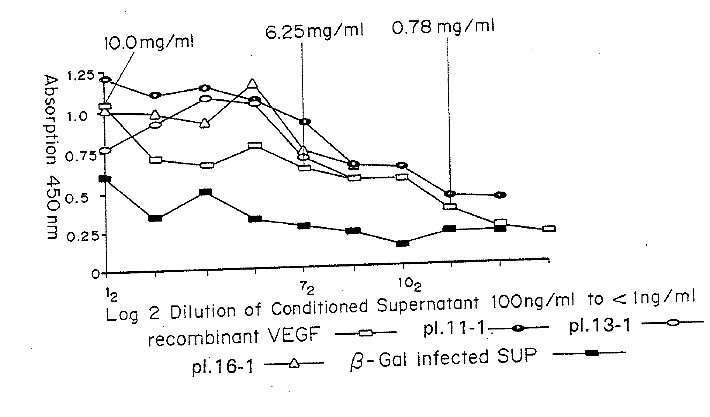
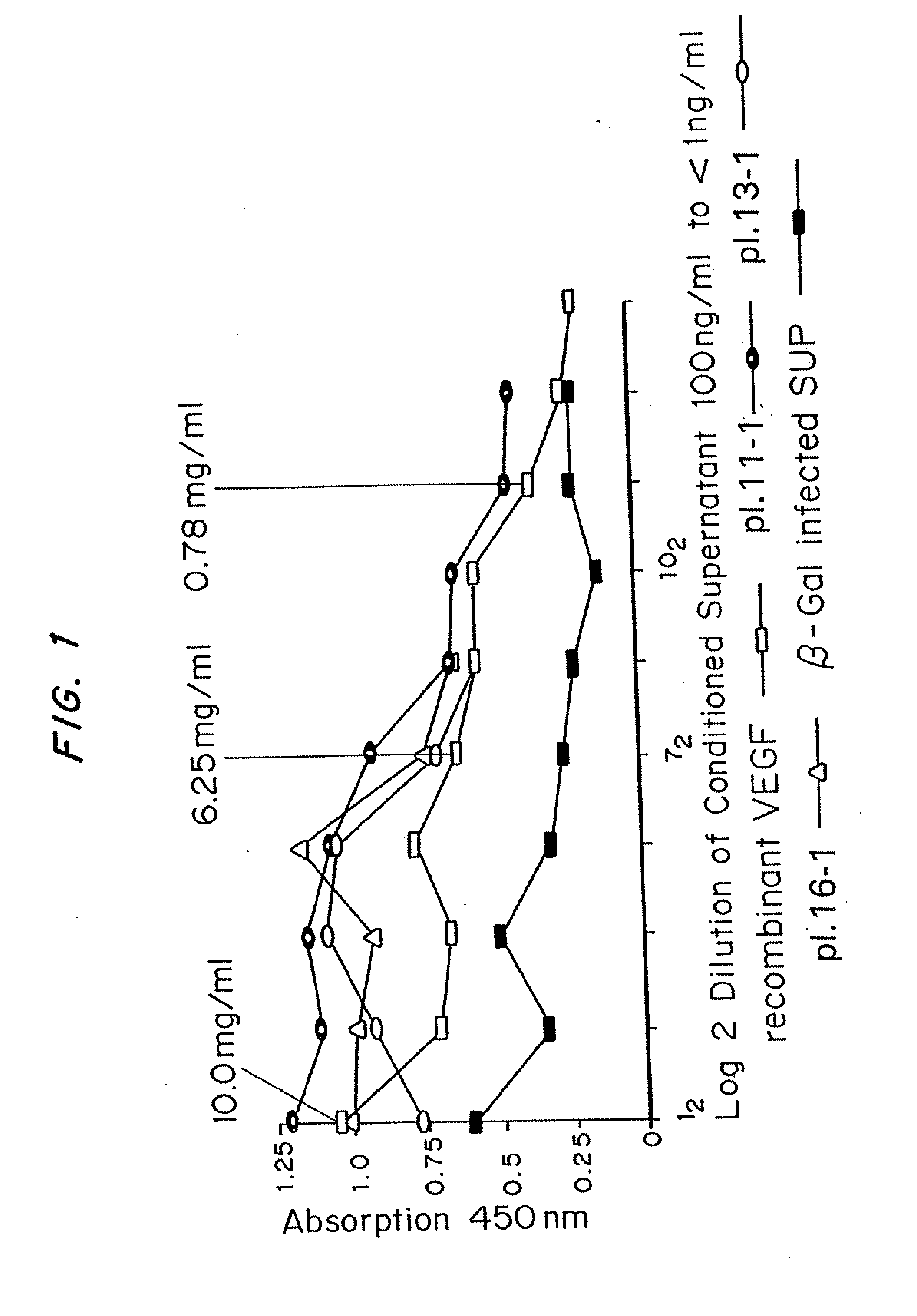
[0053]The human cDNA for VEGF was cloned into a recombinant adenovirus vector. Human umbilical vein endothelial cells (HUVEC) were harvested from fresh umbilical cords using a 0.2% collagenase solution in Hanks Balanced Salt Solution (HBSS) for 20 minutes at room temperature. The cells were washed with HBSS and plated on collagen-coated (1% in PBS) tissue culture dishes in M13 medium supplemented with 20% FBS and 1 mg / ml of bFGF, HUVEC from a confluent 10-cm plate were homogenized and total RNA was extracted with the RNEASY™ kit from QIAGEN® Inc. First strand of cDNA was amplified from the RNA by RT-PCR with oligo-dT primers using the SUPERSCRIPT™ II RT PCR kit from LIFE TECHNOLOGIES®. The full-length human VEGF cDNA was then amplified by PCR with appropriate primers (sense 5′-CCCAAGCTTGCCGCCGCCATGAACTTTC TGCTGTCT-3′ (SEQ ID NO:1); Hind III linker antisense, 5′-GCTCTAGAATCTGGTTCCCGAAACCCTGA-3′ (SEQ ID NO:2), Xba I linker) using pfu (plaque forming units), DNA polymer...

example 3

ADV-VEGF Leads to Angiogenesis

[0062]Mice received subcutaneous injections of ADV-VEGF in one thigh and ADV.LacZ in the other. At various time intervals, these mice were sacrificed and perfusion-fixed. The thighs were harvested and processed for histology and immunohistochemistry. Histology revealed no significant changes in the control (ADV.LacZ)P group and marked presence of new blood vessels in the ADV-VEGF group.

[0063]Thus, it was established that ADV-VEGF infected cells secreted human VEGF165 and that this protein product stimulated endothelial cell proliferation in vitro and angiogenesis in vivo, without local necrosis.

[0064]Examples 4-7 use a mouse model. For all mouse wound experiments, mice were purchased from Jackson Laboratories, and housed one per cage. Animals were anesthetized in a chamber in preparation for all experiments and shaved the day before experimentation. Full thickness wounds were made with a template of 0.8 or 1.4 cm in diameter. All wounds were created in ...


PUM

PropertyMeasurementUnit
Fractionaaaaaaaaaa
Timeaaaaaaaaaa
Timeaaaaaaaaaa

Abstract

A method and means have been developed to deliver a therapeutic dose or dosages of the angiogenic molecule, Vascular Endothelial Growth Factor (VEGF) that results in a statically significant decrease in the time to achieve 100% wound closure and accelerates the rate of healing in experimental diabetic ulcers. Toxicity is evaluated by measuring any local inflammatory response at the wound site, the systemic absorption of VEGF, and the effect on distant organs that may be particularly susceptible to VEGF therapy (e.g., retinopathy and hepatitis) The angiogenic response is quantified by measuring the change in collagen deposition, epithelialization, and the closure rates of diabetic ulcers after therapeutic dosing with ADV-VEGF. Sustained administration of VEGF, stimulates and accelerates the healing process as evidenced by a reduced time to complete healing (defined by 100% epithelialization and no drainage) in experimental diabetic ulcers, with minimal to no toxicity. Important features of the method and reagents for use therein are that the VEGF is released into the ulcer in a sufficient quantity over a period of time for at least two to six weeks, or to closure of the wound.

Description

[0001]This application claims priority to U.S. Ser. No. 60 / 363,584 filed Mar. 12, 2002.[0002]The United States government has certain rights in this invention by virtue of a NIH, National Institute of Diabetes and Digestive and Kidney Diseases (NIDDK) grant R21DK060214-01 and NIH, National Institute of Diabetes and Digestive and Kidney Diseases (NIDDK) grant K08DK059424-01 to Dr. Harold Brem.BACKGROUND OF THE INVENTION[0003]The present invention is a method of tissue engineering, and specifically relates to administration of angiogenic factors directly to diabetic wound ulcers, in an effective amount for a sustained period of time effective to promote closure.[0004]Among the 16 million diabetic patients (diagnosed and undiagnosed) in the United States, an estimated 1200 amputations are performed each week (Pecoraro, et al., Diabetes Care. 1990; 13:213-521); 84% of which are preceded by a foot ulcer. Limb amputation in diabetics is associated with an increased risk for further amputa...

Claims


Application Information

Patent Timeline
IPC IPC(8): A61K48/00A61K38/18A61P7/12A61K31/7088
CPCA61K38/1866A61K48/00A61P7/12
Inventor BREM, HAROLD
Owner NEW YORK UNIV
Why Eureka
Try Eureka