Microstream injector
a technology of injector and microstream, which is applied in the direction of microneedles, infusion needles, other medical devices, etc., can solve the problems of tissue damage, pain in the use of needles, and health workers and patients at risk of infection through inadvertent needle-stick or equipment misuse, so as to discourage accidental contact of spent devices and quickly distinguish the
- Summary
- Abstract
- Description
- Claims
- Application Information
AI Technical Summary
Benefits of technology
Problems solved by technology
Method used
Image
Examples
example 1
Microstream Injection Studies with Gel Models of Tissue
[0071] Embodiments of the invention are tested in polyacrylamide gel systems that stand in for skin and underlying connective tissue, fat, and muscle. Examples of such systems are described by Schramm-Baxter, Katrencik, and Mitragotri (“Jet injection into polyacrylamide gels: investigation of jet injection mechanics”, J. Biomechanics 37 (2004) 1181-1188), Schramm-Baxter and “Needle-free injections: dependence of jet penetration and dispersion in the skin on jet power”, J. Controlled Release 97 (2004) 527-535), and by Shergold, Fleck, and King (“The penetration of a soft solid by a liquid jet, with application to the administration of a needle-free injection, J. Biomechanics, in press 2005, available to online subscribers). In addition to the use of homogeneous gels, biphasic gels are tested, with a tough outer layer, to mimic the stratum corneum, over a softer layer, to mimic the underlying epidermis, dermis, connective tissue,...
example 2
Computer Simulations of Microstream Injections
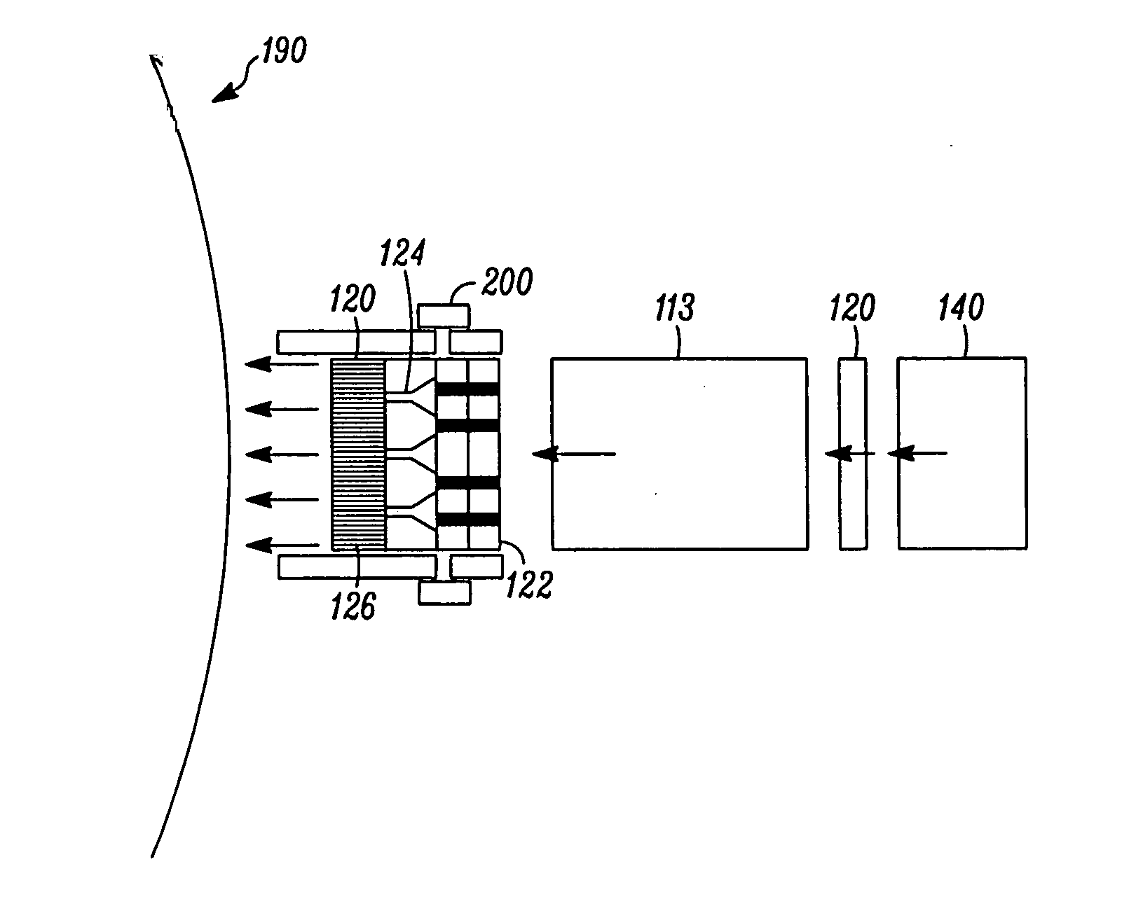
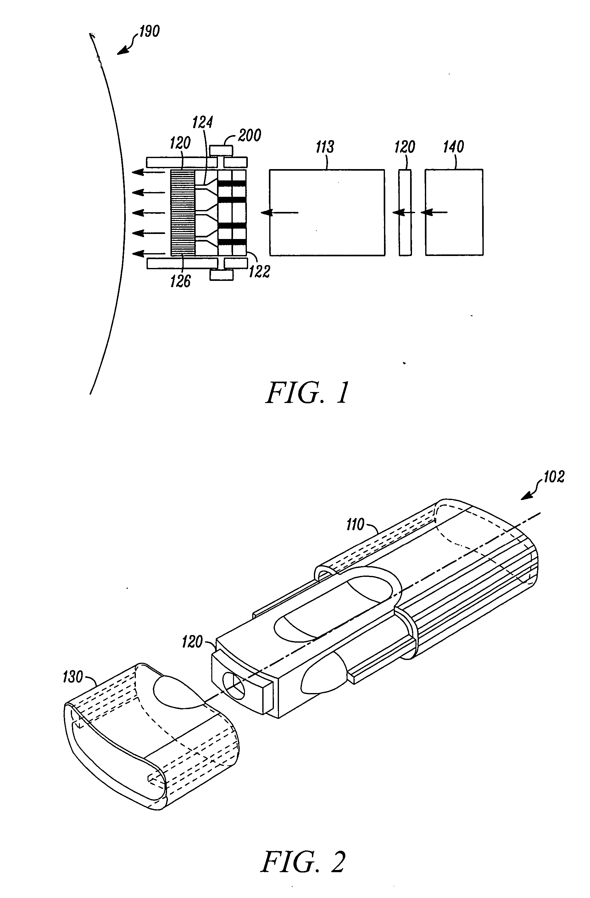
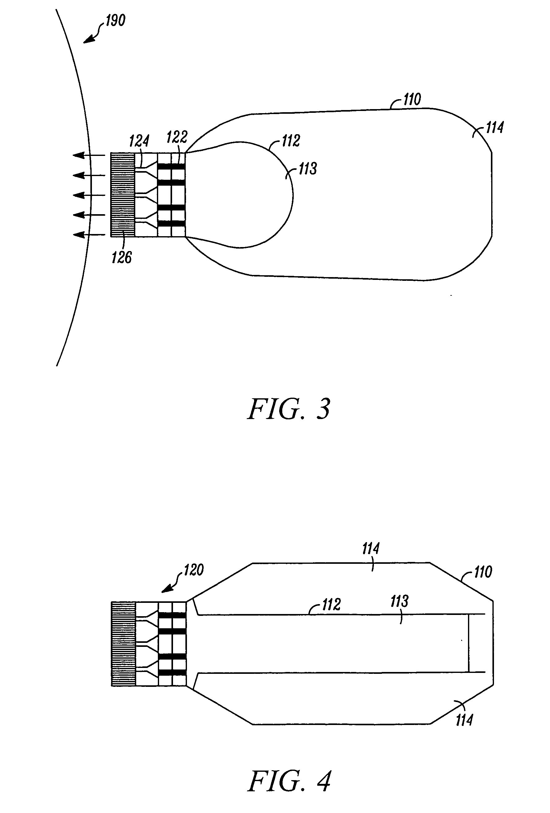
[0072] Computer simulation provides another approach to designing and testing the injector. The inventor has made use of ASE+, a program designed by NASA Marshall Flight Center and the CFD Research Corporation (Huntsville, Ala.), now owned by ESI Corportation (headquarters in Portland Oreg.). As a test model, a nozzle with an upstream width of 500μ, narrowing to a 60μ funnel end with a convergent angle of 60 degrees, emptying into an ejecting tube of 20μ diameter was tested. According to the simulation, an ejection velocity of 60 m / sec is supported by a pressure upstream of the ejecting tube of 3.9 MPa, a velocity of 80 m / sec is achieved with 6.8 MPa, and a 100 m / sec velocity is supported by a pressure of 10.6 MPA. This amount of pressure compares favorably with conventional pressure systems, a CO2 gas cartridge, for example, is filled to a presure of about 7 MPa. In addition to the data supporting the feasibility of attaining appropria...
example 3
In Vitro Transdermal Microstream Injection Studies
[0073] Rates of transdermal transport are determined by contacting human cadaver epidermis with the injector of the invention and activating the device. Skin is obtained from the abdomen or back of human cadavers (with approval of the appropriate Institutional Review Boards). Epidermis is isolated by incubating in 60° C. water for 2 min and gently removing epidermis tissue. Because the primary barrier to transdermal transport is the stratum corneum (the upper 10-15 μm of the epidermis), the use of epidermis rather than full-thickness skin is a well established model for transdermal drug delivery.
[0074] Isolated epidermis is mounted in a Franz diffusion chamber at 37° C. and is bathed in the receiver compartment (lower, viable epidermis side) with PBS, and in the donor compartment (upper, stratum corneum side) with 1 mM calcein (Sigma), 100 units / ml insulin (Humulin-R, Eli Lilly), 80 uM Texas red-labeled BSA (Molecular Probes), or p...
PUM
 Abstract
Description
Claims
Application Information
 - R&D
- Intellectual Property
- Life Sciences
- Materials
- Tech Scout
- Unparalleled Data Quality
- Higher Quality Content
- 60% Fewer Hallucinations
Browse by: Latest US Patents, China's latest patents, Technical Efficacy Thesaurus, Application Domain, Technology Topic, Popular Technical Reports.
© 2025 PatSnap. All rights reserved.Legal|Privacy policy|Modern Slavery Act Transparency Statement|Sitemap|About US| Contact US: help@patsnap.com


