Methods and compositions for pathogen detection using fluorescent polymer sensors
a technology of fluorescent polymer sensors and compositions, applied in the direction of biochemistry apparatus and processes, instruments, material analysis, etc., can solve the problems of time-consuming, difficult to detect pathogens, and difficult to detect food contamination before consumption, etc., to achieve the effect of fast processing and analysis of data and easy preparation
- Summary
- Abstract
- Description
- Claims
- Application Information
AI Technical Summary
Benefits of technology
Problems solved by technology
Method used
Image
Examples
example 1
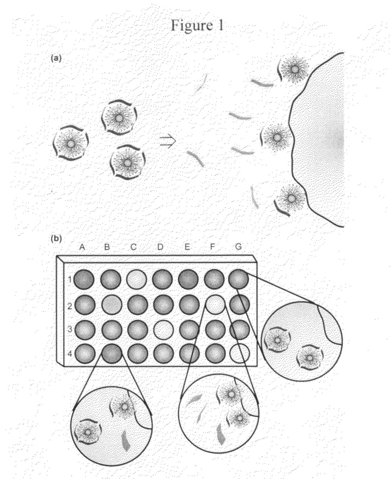
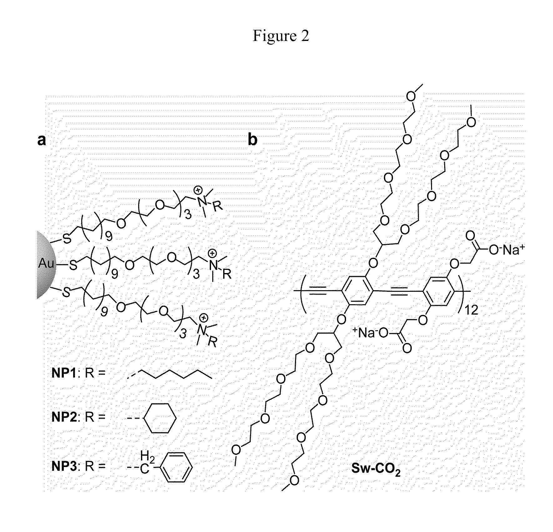
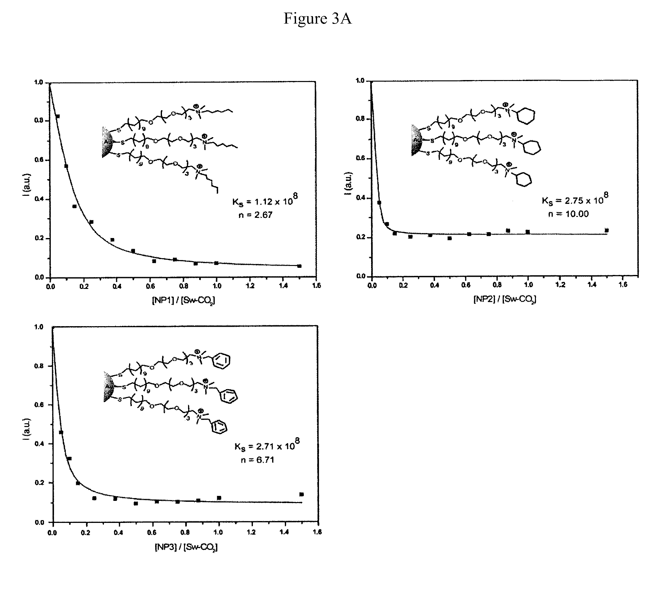
[0037]Fluorescence titration experiments were conducted to evaluate the complexation between nanoparticles and Sw-CO2. For recording the fluorescence response patterns in the presence of bacteria, Sw-CO2 and stoichiometric amounts of NP1-NP3, as determined by the fluorescence titration study (Table 1), were diluted with phosphate buffer (5 mM, pH 7.4) to yield solutions with a final polymer (Sw-CO2) concentration of 100 nM. Subsequently, each solution (200 μL) was placed into a respective well on the microplate. After incubation for 15 min, the fluorescence intensity at 463 nm was recorded with an excitation wavelength of 400 nm.
TABLE 1Binding constants (Ks) and binding stoichiometries (n) betweenanionic polymer (Sw-CO2) and three cationic nanoparticles(NP1-NP3) as determined from fluorescence titration.NanoparticleKs / 108 M−1−ΔG / kJ mol−1nNP11.1245.92.67NP22.7548.110.0NP32.7148.16.71
example 2
[0038]Next, 10 μL of a bacterial solution (OD600=0.05) was added to each well. After incubation for another 15 min, the fluorescence intensity at 463 nm was measured again. The fluorescence intensity before addition of bacteria was subtracted from that obtained after addition of bacteria, to record the overall fluorescence response (ΔI). This process was completed for 12 bacteria to generate six replicates of each, leading to a training data matrix of 3 constructs×12 bacteria×6 replicates (Table 2) that was subjected to a classical linear discriminant analysis (LDA) using SYSTAT (version 11.0). The Mahalanobis distances of each individual pattern to the centroid of each group in a multidimensional space were calculated and the case was assigned to the group with the shortest Mahalanobis distance.
TABLE 2Training matrix of fluorescence response patterns generatedfrom NP-(Sw-CO2) sensor array (NP1-NP3) against varioustypes of bacteria (OD = 0.05 at 600 nm).BacteriaNP1NP2NP3A. azurea1.3...
example 3
[0039]A similar procedure was also performed to identify 64 randomly selected bacterial samples based on their fluorescence response patterns. The classification of new cases was achieved by computing their shortest Mahalanobis distances to the groups generated through the training matrix (3 constructs (NP1-NP3)×12 bacteria×6 replicates). During the identification of unknown bacteria, the bacterial samples were randomly selected from the 12 respective bacteria and the solution preparation, data collection, and LDA analysis were each performed by different researchers, resulting in a double-blind process.
TABLE 4Identification of 64 unknown bacterial samples with LDAusing assemblies of Sw-CO2 and NP1-NP3. From the unknown bacterialsamples, 61 out of 64 were correctly identified, resulting in an accuracy of 95.3%.Fluorescenceresponse patternLDA IdentificationCorrect IdentificationEntryNP1NP2NP3BacteriaYes / NO1142.53375.38892.108P. putidaYES2182.858127.943149.733B. lichenformisYES377.468...
PUM
| Property | Measurement | Unit |
|---|---|---|
| excitation wavelength | aaaaa | aaaaa |
| excitation wavelength | aaaaa | aaaaa |
| core diameter | aaaaa | aaaaa |
Abstract
Description
Claims
Application Information
 - R&D
- Intellectual Property
- Life Sciences
- Materials
- Tech Scout
- Unparalleled Data Quality
- Higher Quality Content
- 60% Fewer Hallucinations
Browse by: Latest US Patents, China's latest patents, Technical Efficacy Thesaurus, Application Domain, Technology Topic, Popular Technical Reports.
© 2025 PatSnap. All rights reserved.Legal|Privacy policy|Modern Slavery Act Transparency Statement|Sitemap|About US| Contact US: help@patsnap.com


