Rapid microbial detection and antimicrobial susceptibility testing
a technology of microbial susceptibility testing and microbial detection, which is applied in the field of rapid microbial detection and antimicrobial susceptibility testing, can solve the problems of not being able to provide the attending physician with specific diagnostic information, unable to significantly improve the outcomes of approximately 87% of patients so treated, and not being able to change from ineffective to effective therapy.
- Summary
- Abstract
- Description
- Claims
- Application Information
AI Technical Summary
Benefits of technology
Problems solved by technology
Method used
Image
Examples
example 1
Electrokinetic Concentration
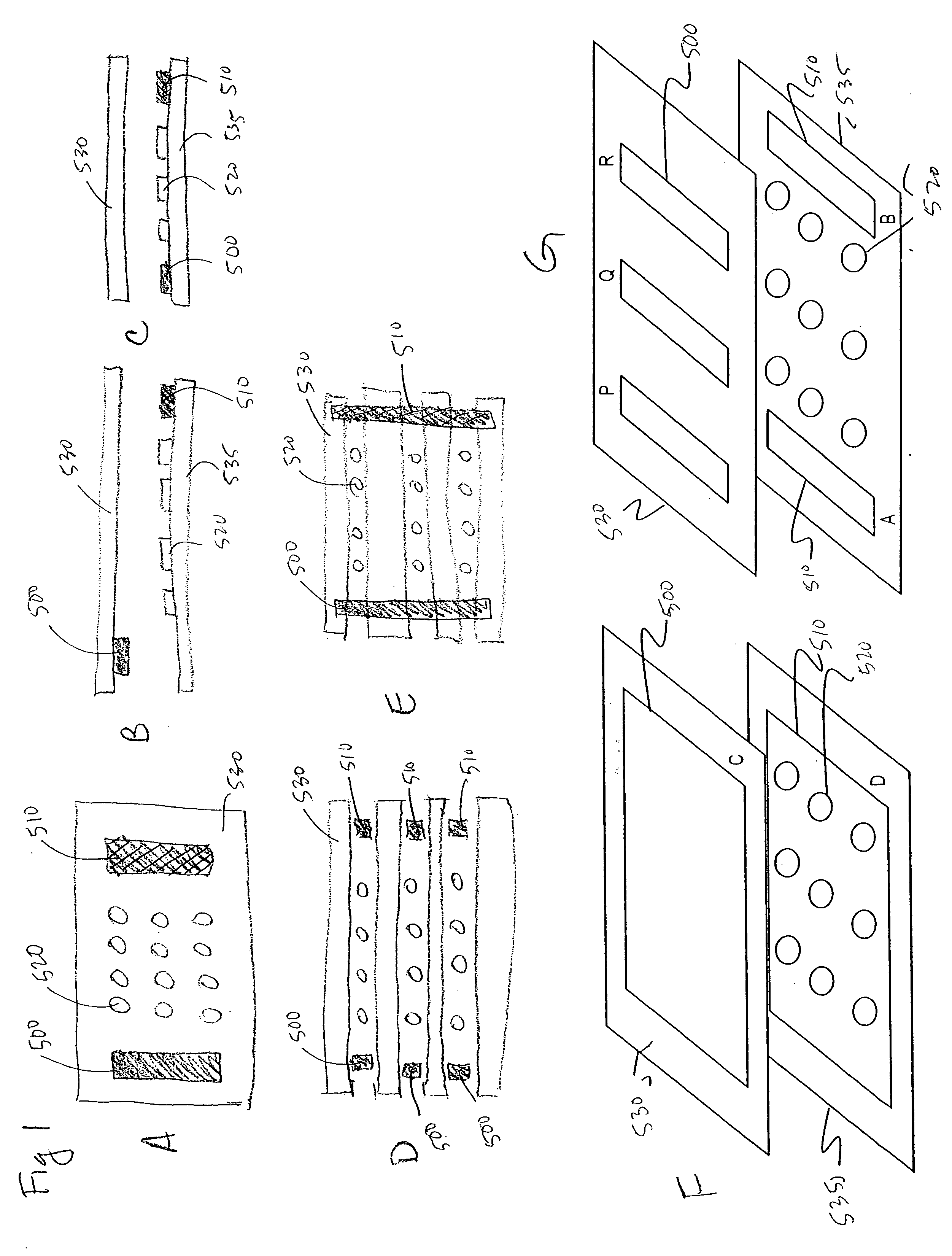
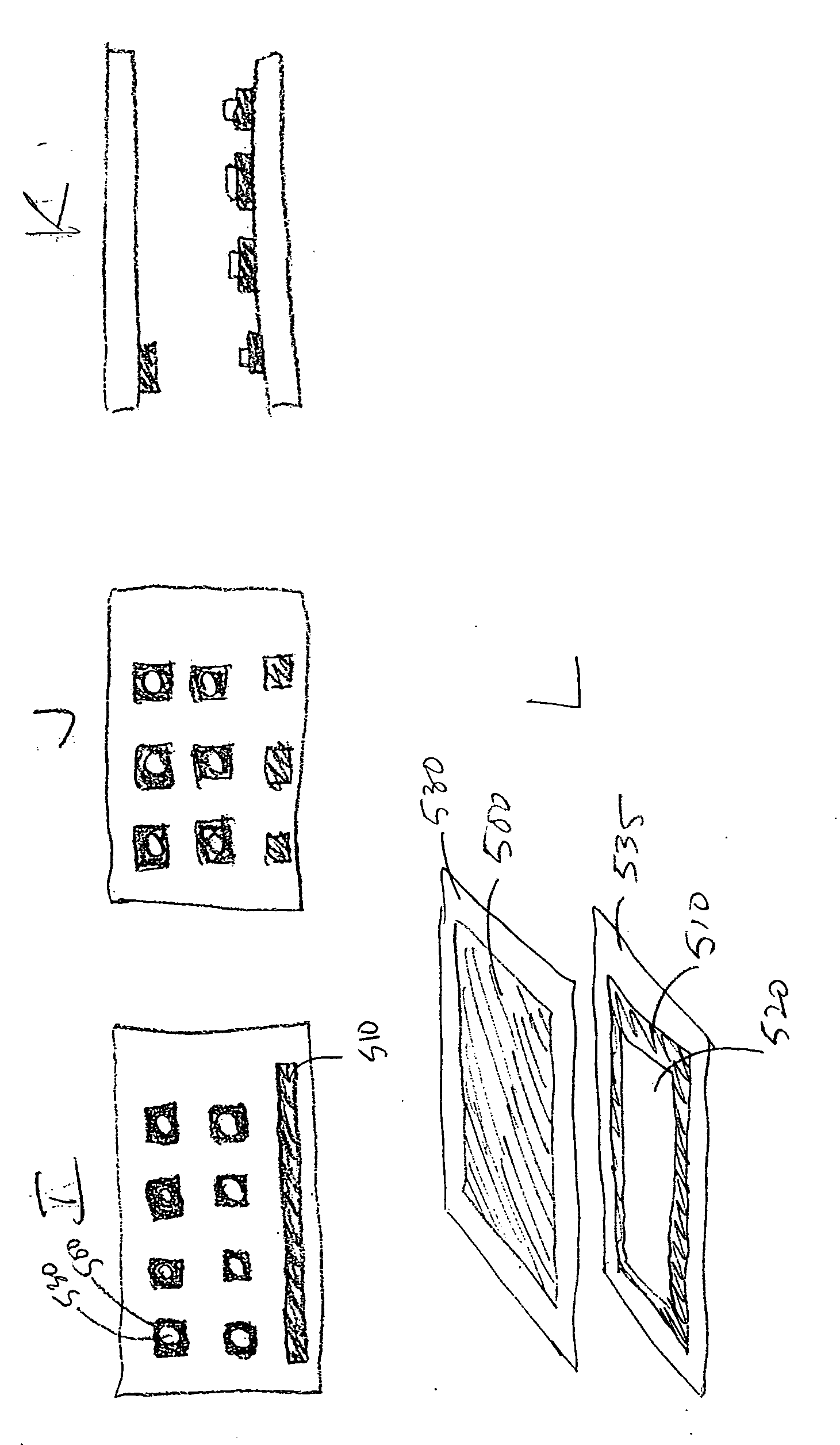
[0322] A flow cell was constructed by sandwiching two ITO modified glass microscope slides between a plastic laminate structure forming a 4 channel flow cell as illustrated in FIG. 50. The distance between the slide surfaces is approximately 500 microns. The bottom ITO slide was precoated with amine reactive OptiChem (Accelr8) and chemically modified with diethylene triamine (DETA) to yield a cationic bacteria capture surface.
[0323] A mixture of 1×10⁷ CFU / ml of Staphylococcus aureus in 10 mM hydroquinone, 10 mM DTT, and 10 mM CAPS pH 6.5 was injected into a flow chamber. A potential difference (1.6 V) was applied across the ITO slides for approximately 3 minutes to surface concentrate and bind the bacteria to the DETA OptiChem modified ITO electrode. Photomicrographs of the surface of the anode before the application of the potential and 50 seconds later show the presence of the electrokinetically concentrated S. aureus microorganisms. The number of bac...
example 2
Microorganism Growth
[0324] The redox solution from Example 1 was replaced with tryptic soy growth media and incubated at 37 C on an inverted microscope to establish detectable growth of bacteria bound to DETA surface. Photomicrographs of the S. aureus at the beginning of growth and after different incubation times were taken (data not shown). The photomicrographs were taken with phase optics, and the flare surrounding the bacteria indicated the presence of additional bacteria resulting from cell division of the original bacteria. Thus bacterial growth ca follow electrokinetic concentration.
example 3
AOA Susceptibility
[0325] Bacteria concentrated and grown as in Examples 1 and 2 were challenged by 5 micrograms / ml of oxacillin to which the S. aureus strain used was susceptible. Simultaneous with the administration of oxacillin was the administration of 10 micromolar propidium iodide, which is excluded from live cells, and thus whose presence in fluorescence images is indicative of cell death. Photomicrographs of the bacteria at the beginning of AOA treatment were taken, under phase contrast and fluorescence microscopy, and additional images were taken one hour later (data not shown). There was a large increase in fluorescence which indicated the large amount of cell death from the administration of oxacillin.
Example 4
Modeling of AOA Susceptibility
[0326] The effect of oxacillin on S aureus can be modeled using a mathematical Hill function: Effect=Emax×tnET50+tn
[0327] Where Emax is equivalent to the maximum effect over time t, ETW is the time at which half the maximal effect i...
PUM
 Abstract
Description
Claims
Application Information
 - R&D
- Intellectual Property
- Life Sciences
- Materials
- Tech Scout
- Unparalleled Data Quality
- Higher Quality Content
- 60% Fewer Hallucinations
Browse by: Latest US Patents, China's latest patents, Technical Efficacy Thesaurus, Application Domain, Technology Topic, Popular Technical Reports.
© 2025 PatSnap. All rights reserved.Legal|Privacy policy|Modern Slavery Act Transparency Statement|Sitemap|About US| Contact US: help@patsnap.com


