
Bone and/or joint disease-associated genes

a technology of gene and bone, applied in the field of bone and/or joint disease-associated genes, can solve the problems of difficult identification of genes, inability to identify genes by itself, and lack of medical devices having a mechanism that clearly inhibits cartilage degeneration and stimulates cartilage regeneration. to achieve the effect of stimulating cartilage differentiation and increasing or decreasing expression levels

Inactive Publication Date: 2007-02-01
KOMORI TOSHIHISA +1
View PDF0 Cites 20 Cited by
  • Summary
  • Abstract
  • Description
  • Claims
  • Application Information

AI Technical Summary

Benefits of technology

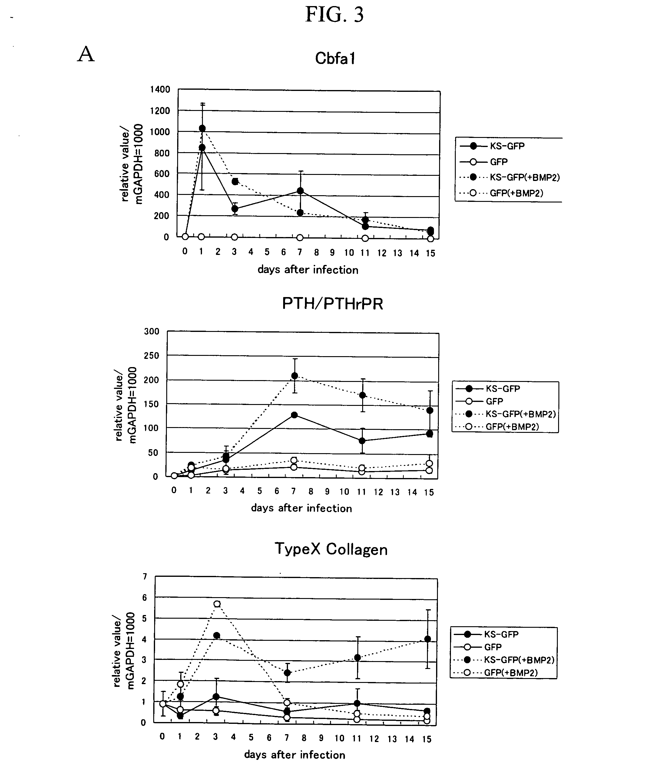
[0020] The second aspect of the present invention provides primary chondrocytes derived from a Runx2 / Cbfa1-deficient mouse or cultured chondrocytes and chondrocytes derived from a Runx2 / Cbfa1- and p53-deficient mouse that are employed in the first aspect of the present invention. More specifically, the method for obtaining genes using such cells according to the first aspect of the present invention does not induce constitutive gene expression by endogenous Cbfa1 because of deficiency in Cbfa1 transcription factors and can provide a system for inducing and / or inhibiting gene expression with lower background levels and higher detection sensitivity compared with conventional techniques. The Runx2 / Cbfa1- and p53-deficient mouse-derived chondrocyte cell lines that can be preferably used in the present invention are deposited as the RU-1 and RU-22 cell lines at the International Patent Organism Depositary of the National Institute of Advanced Industrial Science and Technology (Tsukuba Central 6, 1-1-1 Higashi, Tsukuba, Ibaraki, 305-8566, Japan) under the Budapest Treaty as of Aug. 5, 2003, under the accession numbers FERM BP-10137 (the RU-1 cell line) and FERM BP-10138 (the RU-22 cell line) (the original deposit).
[0023] The aforementioned downstream gene of Runx2 / Cbfa1 is derived from a mouse. By searching the database, a human homologue thereof can be easily identified, and it can be easily deduced that such human homologue has similar effects concerning cartilage differentiation. Based on such genetic information, homologous genes can be easily obtained by hybridization. Thus, it can be easily deduced that the resulting gene has similar functions concerning cartilages. Accordingly, the present invention also provides a human-derived polynucleotide, which is homologous to the aforementioned mouse-derived polynucleotide having the nucleotide sequence shown in SEQ ID NO: 27, 29, 31, 33, 35, 37, 39, 41, 43, 45, 47, 49, or 51.
[0027] The downstream gene of Runx2 / Cbfa1, which had been found to inhibit cartilage differentiation by the aforementioned method, can inhibit the stimulated cartilage differentiation observed in a bone and / or joint disease (preferably osteoarthritis) with the use of a polynucleotide of the gene, a polypeptide encoded by such gene, a vector containing the polynucleotide of the gene, a compound that activates the gene expression or protein functions, or the like. Thus, therapeutic and / or prophylactic effects on a bone and / or joint disease can be expected. In contrast, the downstream gene of Runx2 / Cbfa1, which had been found to stimulate cartilage differentiation, can inhibit the stimulated cartilage differentiation observed in a bone and / or joint disease (preferably osteoarthritis) with the use of a compound that inhibits the protein functions, a compound that inhibits the gene expression, an antibody, an RNAi molecule, an antisense polynucleotide, or the like. Thus, therapeutic and / or prophylactic effects on a bone and / or joint disease can be expected.

Problems solved by technology

However, no medicine having a mechanism that clearly inhibits cartilage degeneration and stimulates cartilage regeneration has yet been developed.
Accordingly, it is deemed difficult to identify genes that play a key role in the advancement of disease via simple comparison between pathologic tissues and normal tissues.
Since an inflammatory cytokine also has enormous numbers of functions, this technique by itself cannot successfully identify the genes that play a key role in inflammation or disease.
Accordingly, a medicine that targets the direct inhibition of Runx2 / Cbfa1 functions may cause side effects.
However, thorough analysis of the downstream genes of Runx2 / Cbfa1has not been attempted in the past.
Thus, it may be difficult to detect all the NF-kB-regulated genes with high sensitivity.
A method that can overcome such difficulty is deemed necessary, although such method has not yet been developed.

Method used


Image

  • Bone and/or joint disease-associated genes
  • Bone and/or joint disease-associated genes
  • Bone and/or joint disease-associated genes

Examples


example 1

Isolation of Primary Chondrocyte from Runx2 / Cbfa1-Deficient Mouse Fetus and Establishment of Chondrocyte Cell Lines from Runx2 / Cbfa1- and p53-Deficient Murine Embryonic Skeleton

(1) Sampling of Primary Chondrocyte Derived from Runx2 / Cbfa1− / − Mouse

[0217] The Runx2 / Cbfa1 knockout mice prepared by Komori et al. were used (Cell, 1997, 89, 755-764, JP Patent Publication (Unexamined) No. 10-309148 (1998)). The skeletons were collected from the Runx2 / Cbfa1-homodeficient mice at day 18.5 of embryonic development, and the skeletons were treated with a solution containing 0.1% EDTA and 0.1% Trypsin (pH 7.4) at 37° C. for 60 minutes. Thereafter, the skeletons were treated with 1.5 mg / ml collagenase and alpha modified-minimum essential medium (αMEM) for 3.5 hours to obtain a cell suspension. The cell suspension was cultured on a collagen-coated dish containing Dulbecco's Modified Eagle's Medium / Ham's F12 (1:1) hybrid medium (Gibco BRL) containing 10% fetal bovine serum for 2 or 3 days for cel...

example 2

Construction of a System of Inducing Cartilage Differentiation via Forced Expression of Runx2 / Cbfa1 Using an Adenovirus

(1) Construction of an Adenovirus for the Expression of Runx2 / Cbfa1

[0221] cDNA comprising the open reading frame (ORF) of murine Runx2 / Cbfa1 was inserted into the BarnHI site of pIRES2-EGFP (Biosciences Clontech), and a fragment containing Runx2 / Cbfa1, the internal ribosome entry site (IRES), and the enhanced green fluorescence protein (EGFP) was inserted into the BamHI-XbaI site of the shuttle vector pACCMV.pLpA (Proc. NatI. Acad. Sci. U.S.A., 1993, 90, 2812-2816). The constructed vector was cotransfected into the human kidney cell line 293 with the adenovirus vector pJMI7 (Virology, 1988, 163, 614-617) using the Superfect transfection reagent (Qiagen). The adenovirus retaining a Runx2 / Cbfa1 fragment resulting via homologous recombination was subjected to subculture 3 or 4 times with the use of the cell line 293 to multiply the viruses. A roughly purified stock ...

example 3

Isolation of Downstream Gene of Runx2 / Cbfa1 via Subtraction

[0228] The downstream gene of Runx2 / Cbfa1 was obtained via subtraction using the undifferentiated mesenchymal cell line (C3H10T1 / 2). At the outset, the C3H10T1 / 2 cell line that intensively expresses Runx2 / Cbfa1 was established (C3H10T1 / 2-Runx2 / Cbfa1). Subsequently, C3H10T1 / 2-Runx2 / Cbfa1 and C3H10T1 / 2 were used to screen for the gene that is intensively expressed in the C3H10T1 / 2-Runx2 / Cbfa1 cell line via subtraction. Subtraction was carried out using the CLONTECH PCR-Select™ cDNA Subtraction Kit in accordance with the user's manual. As a result, the expression level of the gene shown in SEQ ID NO: 11 (Riken cDNA 2810002E22 gene (HNOEL-iso homolog)) was found to be higher in C3H10T1 / 2-Runx2 / Cbfa1 than in C3H10T1 / 2.

[0229] Whether or not the HNOEL-iso homolog gene is induced by Runx2 / Cbfa1 was analyzed via real-time PCR in RU-1, RU-22, and Runx2 / Cbfa1− / −mouse-derived primary cultured chondrocytes, in order to confirm that the...


PUM


Abstract

This invention provides a method for obtaining a gene involved in regulation of cartilage differentiation, in which a transcription factor, preferably Runx2 / Cbfa1, is forcedly expressed in a cell that is deficient such transcription factor, preferably in a Runx2 / Cbfa1-deficient chondrocyte, and the gene, the expression of which is thereby induced, is selected using DNA chip analysis, subtraction, or other means as well as a Runx2 / Cbfa1-deficient chondrocyte useful for carrying out such method. The invention also provides a polynucleotide obtained by such method, a polypeptide encoded by such polynucleotide, an antibody against such polypeptide, a recombinant vector comprising such polynucleotide, a transformant comprising such recombinant DNA vector, a cell expressing such polypeptide, a transgenic animal of such polynucleotide, an animal model of a bone and / or joint disease (preferably osteoarthritis), a method for screening for a therapeutic agent and / or prophylactic agent for a bone and / or joint disease (preferably osteoarthritis) using the aforementioned objects, a candidate compound for a therapeutic agent and / or prophylactic agent selected by such method, a pharmaceutical composition for a bone and / or joint disease (preferably osteoarthritis), and a method for diagnosing such disease.

Description

TECHNICAL FIELD [0001] The present invention relates to proteins having a novel function of regulating cartilage differentiation, genes encoding such proteins, and a method for isolating such genes. Further, the present invention relates to a method for screening for therapeutic agents for a bone and / or joint disease (preferably osteoarthritis) as well as therapeutic agents for such diseases. BACKGROUND ART [0002] Osteoarthritis (OA), which is a bone and / or joint disease, causes cartilage degeneration and spur formation in the course of aging or receipt of dynamic stress, and OA involves a secondary synovitis. Besides aging, sexuality (female), obesity, and external injuries (such as ligament or medial meniscus injuries) can be risk factors, although causal factors of OA have not been clarified. It is reported that approximately 900,000 people are newly afflicted with osteoarthritis per year in Japan (Non-Patent Document No. 1: Nankodo, Co., Ltd., Orthopedic surgery 42: 2-6, 2002). ...

Claims


Application Information

Patent Timeline
Patent Type & Authority Applications(United States)
IPC IPC(8): A01K67/027C12Q1/68C07H21/04C12N5/06C07K14/705A61K38/00C07K14/47
CPCA61K38/00C12Q2600/158C07K14/47A61P19/00A61P19/02A61P19/08A61P29/00
Inventor KOMORI, TOSHIHISAKANATANI, NAOKOYOSHIDA, CAROLINA ANDREAZANMA, AKIRAKOBAYASHI, SHINJIYAMANA, KEI
Owner KOMORI TOSHIHISA
Learn More