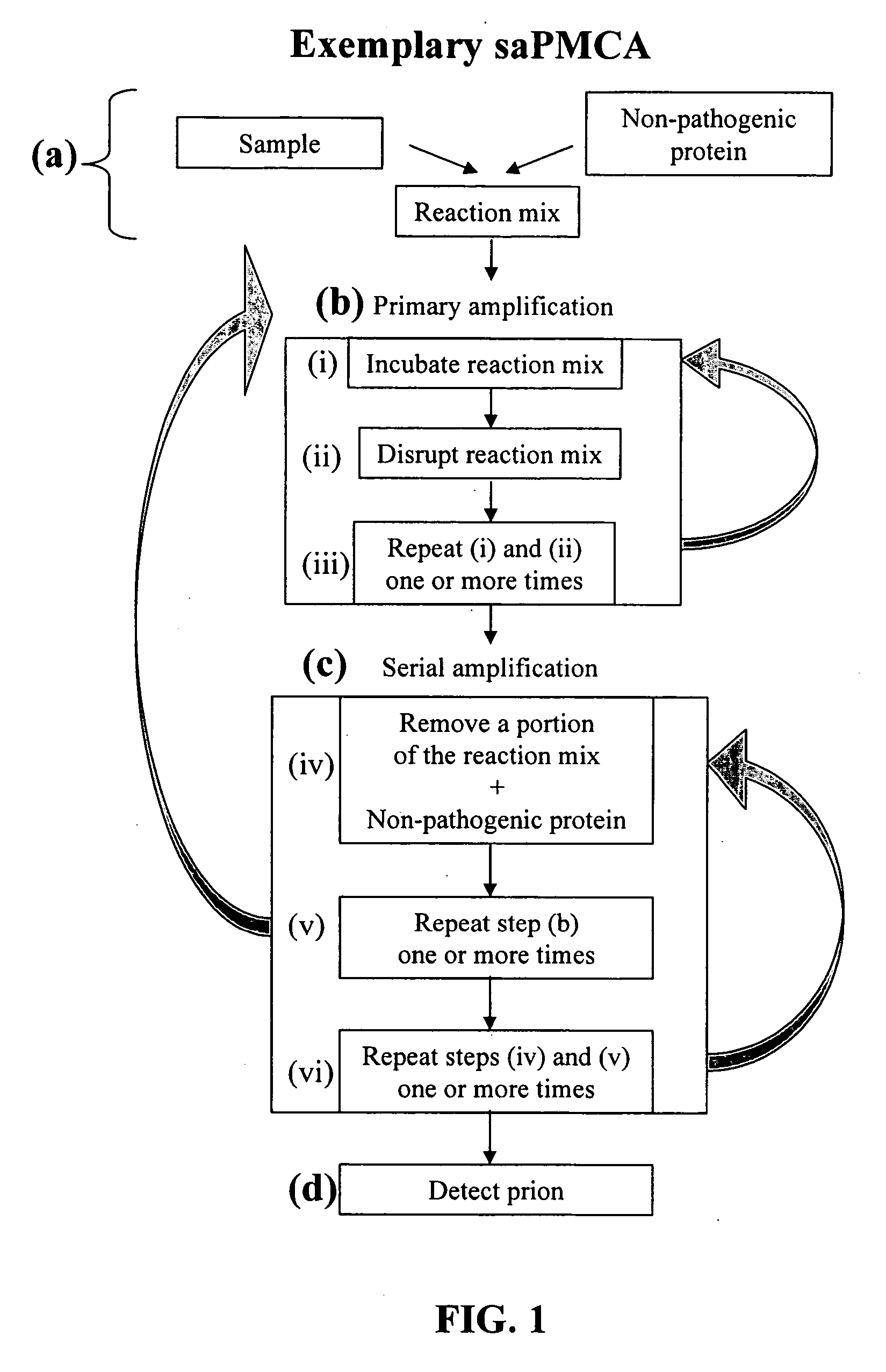
Ultrasensitive detection of prions by automated protein misfolding cyclic amplification
a technology of protein misfolding and cyclic amplification, applied in the field of pathology, biochemistry, cell biology, can solve the problems of inability to carry out large-scale, time-consuming and laborious procedures, and sometimes false-negative brain biopsy results,
- Summary
- Abstract
- Description
- Claims
- Application Information
AI Technical Summary
Benefits of technology
Problems solved by technology
Method used
Image


Examples
example 1
Purification of PrPSc from Brain
[0156] One gram of brain tissue was homogenized in 5 ml of cold PBS containing protease inhibitors. For PMCA-generated PrPres, after the last amplification, the total sample containing the normal brain homogenate used as a substrate was processed in the same way as brain homogenate. The samples were mixed with 1 volume of 20% sarkosyl and the mixture was homogenized and sonicated until a clear preparation was obtained. Samples were centrifuged at 5000 rpm for 15 min at 4° C. The pellet was discarded and the supernatant was mixed with ⅓ volume of PBS containing 0.1% SB-314 and samples were centrifuged in a Biosafe Optima MAX ultracentrifuge (Beckman Coulter, Fullerton, Calif.) at 100,000×g for 3 hr at 4° C. Supernatant was discarded and pellets were resuspended in 600 μl of PBS containing 0.1% SB-314, 10% NaCl and sonicated. The resuspended pellet was layered over 600 ul of PBS containing 20% saccharose, 10% NaCl and 0.1% SB 3-14 and centrifuged for 3...
example 2
Automation: Increase throughput and decrease on Time for Amplification
[0157] The use of a single-probe traditional sonicator imposes a practical problem for handling many samples simultaneously, as a diagnostic test would require. The inventors have adapted the cyclic amplification system to a 96-well format microplate sonicator (the Misonix™ Model 3000 (Farmingdale, N.Y.)), which provides sonication to all the wells at the same time and can be programmed for automatic operation. This improvement not only decreases processing time, increase throughput, and allows performing routinely many more cycles than single-probe sonicator, but also prevents loss of material. Cross contamination is eliminated since there is no direct probe intrusion into the sample. The latter is essential to handle infectious samples and to minimize false positive results. Ten cycles of 1 h incubation followed by sonication pulses of 30 seconds gave a significant amplification of PrPSc signal, similar to that...
example 3
Increase on Amplification Efficiency by Metal Chelation
[0159] As part of our efforts to optimize the PMCA procedure the inventors discovered that the relatively low level of amplification observed previously was in part due to the presence of metal cations in the samples. When the PMCA reactions were done in the presence of EDTA, a broad-range metal chelator, the efficiency of amplification was dramatically higher.
[0160] It is well established that PrP binds copper (and to a lesser extent zinc) with high affinity and indeed a possible biological function for the normal prion protein is to participate in Cu2+ transport across the cell membrane (Brown and Sassoon, 2002). The results indicate that the positive effect of EDTA in boosting PMCA efficiency was lost when Cu2+ was added to the reaction. The effect is very clear and is concentration-dependent. No significant effect was observed with other divalent cations such as Ca2+ and Mg2+, but Zn2+ also decreased efficiency of prion co...
PUM
| Property | Measurement | Unit |
|---|---|---|
| Temperature | aaaaa | aaaaa |
| Time | aaaaa | aaaaa |
| Time | aaaaa | aaaaa |
Abstract
Description
Claims
Application Information
 - R&D
- Intellectual Property
- Life Sciences
- Materials
- Tech Scout
- Unparalleled Data Quality
- Higher Quality Content
- 60% Fewer Hallucinations
Browse by: Latest US Patents, China's latest patents, Technical Efficacy Thesaurus, Application Domain, Technology Topic, Popular Technical Reports.
© 2025 PatSnap. All rights reserved.Legal|Privacy policy|Modern Slavery Act Transparency Statement|Sitemap|About US| Contact US: help@patsnap.com