
Ophthalmic implant for treatment of glaucoma

a technology of ophthalmological implants and glaucoma, which is applied in the field of ophthalmological implants for treatment of glaucoma, can solve the problems of surgical treatment, limited the ability of the inner wall to be distended outward, and does not teach a way to increase the aqueous outflow by non-drug induced tension, etc., to achieve sufficient tensioning force, increase fluid outflow, and increase fluid permeability

Active Publication Date: 2006-08-31
NOVA EYE INC
View PDF27 Cites 121 Cited by
  • Summary
  • Abstract
  • Description
  • Claims
  • Application Information

AI Technical Summary

Benefits of technology

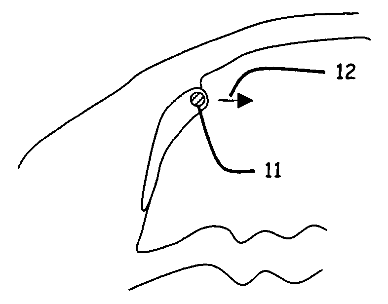
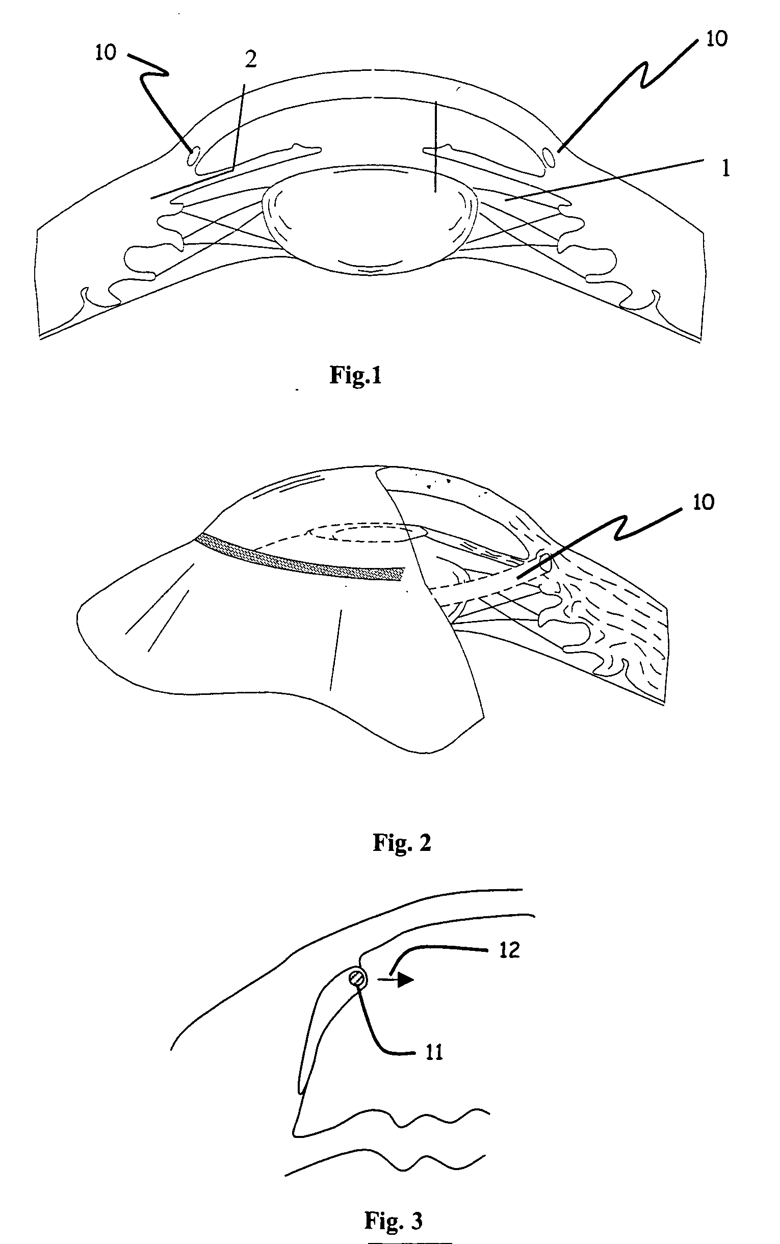
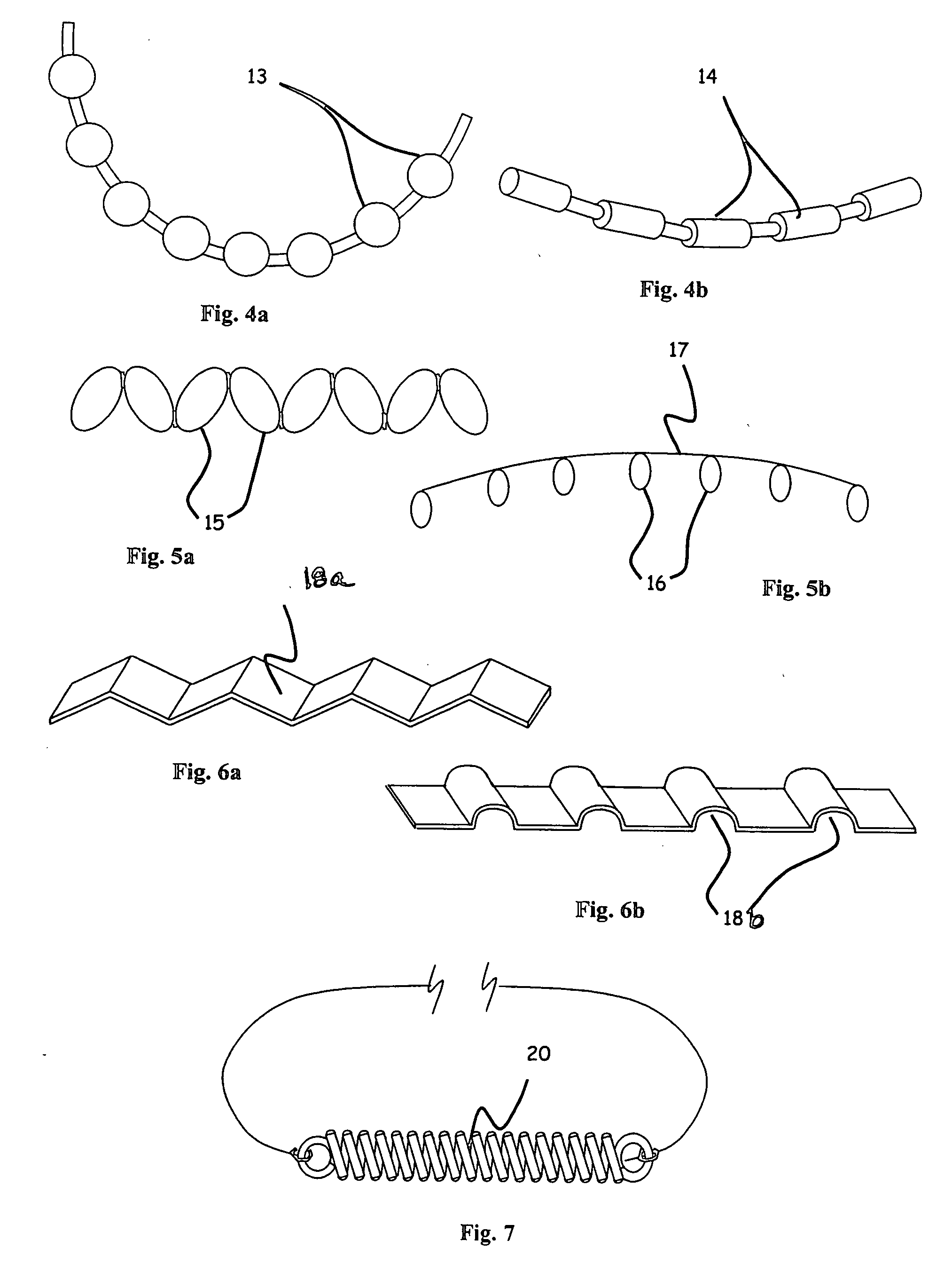
[0020] A method is provided for increasing the outflow of fluid through Schlemm's canal that is useful for treatment of glaucoma. The implant is placed in Schlemm's canal by use of a flexible delivery instrument attached to the implant. The method comprises positioning the instrument and implant within the canal, releasing the implant and connecting the distal and proximal ends of the implant to apply sufficient tensioning force on the inner wall of the canal to increase fluid permeability. A method is also provided for increasing the aqueous outflow of fluid through Schlemm's canal in the eye by positioning in the canal a delivery instrument attached to the implant securing one of the distal or proximal ends of the implant within the canal, adjusting the implant to provide sufficient tensioning force on the inner wall of the canal to increase fluid permeability of the inner wall of the canal, and securing the other of the distal or proximal ends within said canal to maintain the tensioning force on the inner wall of the canal.

Problems solved by technology

Such aqueous outflow conditions are usually treated by topical drugs in the form of eye drops, but may result in surgical treatment if drug treatment becomes ineffective due to loss of response to the drug or poor patient compliance.
Post-surgical complications and bleb management are significant issues with trabeculectomy and similar procedures.
In addition, dilation of the canal may also limit the ability of the inner wall to be distended outward by increased intraocular pressure and press against the outer wall to increase aqueous outflow resistance (Ellingsen, B. A., Grant, W. M., Investigative Ophthalmology, 1972; 11(1): 21-8).
While the prior art describes various ways to bypass or to stent the lumen of Schlemm's canal, it does not teach a way to increase aqueous outflow by applying non-drug induced tension to the trabecular meshwork at the inner wall of the canal.

Method used


Image

  • Ophthalmic implant for treatment of glaucoma
  • Ophthalmic implant for treatment of glaucoma
  • Ophthalmic implant for treatment of glaucoma

Examples


example 1

[0058] Human whole globes were obtained from a qualified tissue bank. The eyes were inflated to approximately 10 mm Hg anterior chamber pressure and placed in a cup-shaped holder. A single flap scleral dissection was performed and Schlemm's canal was exposed, allowing access to the ostia of the canal. A 200 micron diameter ophthalmic microcannula (iTRACK™ microcannula, iScience Surgical Corp.) was used to cannulate the entire canal circumference. The microcannula was designed to provide a means of expanding Schlemm's canal through the injection of viscoelastic materials, and comprised an illuminated beacon tip to provide visual indication of the location of the microcannula when in the canal.

[0059] The microcannula was primed with viscoelastic (Healon GV, Advanced Medical Optics, Inc) and inserted into the ostia of the canal. Using a minimal amount of viscoelastic injection to open the canal and provide a lubricious environment, the microcannula was advanced completely around the c...

example 2

[0060] A human whole globe was prepared similar to Example 1 above. The globe was placed in a room temperature bath of phosphate buffered saline, and a 30 gauge infusion needle placed through the cornea to provide fluid input. The needle was attached to a reservoir containing phosphate buffered saline set at a height of 5.4 inches over the height of the Anterior Chamber to provide a constant pressure infusion of 10 mm Hg. The fluid circuit also comprised a digital flow meter (Sensiron, Inc) to record the flow rates on a PC computer. A stabile baseline flow reading was taken prior to surgical cut-down. Surgical access and implant placement as described in Example 1 above was performed, with the exceptions that a two-flap access was used similar to viscocanalostomy and deep sclerectomy surgery, and the filament was not tied. The ends of the filament were brought out from under the surgical flap and the surgical flap was carefully sealed with cyanoacrylate adhesive without adhering the...

example 3

[0062] In another set of experiments, each end of the 28 micron diameter filament implanted in Schlemm's canal was attached to 30 gram mechanical force gauges (Wagner Instruments) in a fixture such that tension forces could be applied to the filament ends. Outflow measurements were taken at three different intraocular pressure values and with varying tension. The value of the Outflow Facility was calculated by dividing the outflow rate in ul / min by the pressure in mm Hg. The experimental results indicate an optimal value for increasing outflow facility in a tension range of approximately 6-10 grams force along the entire length of the implant long axis, or 0.159 to 0.265 grams per mm arc length of canal for a nominal 12 mm diameter of curvature canal. Outflow was significantly increased in the tension range of 1 to 11 grams force or 0.027 to 0.292 grams force per mm of arc length of the canal for a nominal 12 mm diameter of curvature canal. Further increases in tension led to decrea...


PUM


Abstract

An implant is placed within Schlemm's canal of the eye and provides tension to the trabecular meshwork. The tension is continuous and increases the aqueous outflow without the necessity of administering cholinergic drugs to treat glaucoma.

Description

CROSS-REFERENCE TO RELATED APPLICATION [0001] This application claims priority under 35 U.S.C. 119(e) from Provisional U.S. Patent Application Ser. No. 60 / 637,368, filed Dec. 16, 2004 (Atty. Docket No. ISSCP007P), entitled “OPHTHALMIC IMPLANT FOR TREATMENT OF GLAUCOMA”, which is incorporated herein by reference.BACKGROUND OF INVENTION [0002] Glaucoma is a disease condition of the eye in which increased intraocular pressure (IOP) is created by blockage of the drainage mechanism for the aqueous fluid produced in the anterior portion of the eye. Such aqueous outflow conditions are usually treated by topical drugs in the form of eye drops, but may result in surgical treatment if drug treatment becomes ineffective due to loss of response to the drug or poor patient compliance. [0003] Ophthalmic research has indicated that the greatest resistance to aqueous outflow is in the trabecular meshwork, more specifically, the juxtacanalicular tissue at the inner wall of Schlemm's canal (Grant, M....

Claims


Application Information

Patent Timeline
Patent Type & Authority Applications(United States)
IPC IPC(8): A61F2/14
CPCA61F9/00781A61F2/14A61F9/007
Inventor STEGMANN, ROBERTCONSTON, STANLEY R.KUPIECKI, DAVID J.MCKENZIE, JOHNPINSON, CANDICE D.YAMAMOTO, RONALD K.
Owner NOVA EYE INC
Learn More