
Expressing TGF-beta proteins in plant plastids

a technology of plastids and proteins, applied in the field of plastid bioactive tgf proteins, can solve the problems of insufficient production of holo-mis precursors, inability to provide mis at the level required for clinical trials or commercial applications, and inability of bacterial and mammalian systems to directly produce c-terminal mis without a refolding process,

Inactive Publication Date: 2006-07-20
CHLOROGEN
View PDF0 Cites 9 Cited by
  • Summary
  • Abstract
  • Description
  • Claims
  • Application Information

AI Technical Summary

Benefits of technology

[0066] The present invention also relates to plastid transformation vectors and transplastomic plant cells and transplastomic plants, all of which contain a polynucleotide sequence that encodes a full length or truncated TGF-β protein whereby TGF-β protein is expressed in the transgenic plastid / plant and the TGF-β protein thus produced is bioactive, i.e., it binds with its corresponding TGF-β receptor or can be processed to a truncated version that can bind with its receptor. The TGF-β protein can be the full length protein, a bioactive truncated version of the protein, or a modified TGF-β protein. In addition to the polynucleotide expression cassette (coding and regulatory sequences) present in a plastid transformation vector, the vectors will also contain flanking DNA on each side of the polynucleotide expression cassette wherein both flanking sequences are homologous to a DNA sequence of the target plastid genome. The flanking sequences facilitate the stable integration of the expression cassette into the plastid genome through homologous recombination. In a preferred embodiment the plastid transformation vectors contain a selectable marker coding sequence to facilitate the identification of transplastomic plant cells.
[0087] In an alternative embodiment, chloroplast promoters that express the gene product in non-green tissues can be used to express a selectable marker gene so that non-green tissue can be used as a transformation target and selection can begin in culture in non-green tissue. This allows for more flexibility in the regeneration of transformed cells into plantlets, in particular where it is advantageous or necessary to form somatic embryos in order to regenerate a transgenic plant from a transgenic plant cell. (See WO 2004 / 005480 titled “Chloroplast Genetic Engineering Via Somatic Embryogenesis”)
[0090] As a practical matter, the expression cassette will also have a coding sequence for a selectable marker protein. The selectable marker gene can encode, for example, any enzyme that will inactivate a selection agent. Selectable marker genes and selection agents are well known to one of ordinary skill in the art. The selection agent can be a compound that is toxic to the target plant tissue that is being transformed or alternatively it can be a compound that is only toxic to plastids. Selection agents toxic to plants that are known in the art include chloramphenicol, kanamycin, neomycin, basta, glyphosate, etc., as well as their corresponding selectable marker genes, i.e., CAT, bar / PAT, NPTII, EPSPS, etc. Spectinomycin and streptomycin are two selection agents that are only toxic to the plastids. Expression of the aadA gene confers resistance to spectinomycin and streptomycin, and thus allows for the visual identification of plant cells expressing this marker. The aadA gene product allows for continued growth and greening of cells whose chloroplasts comprise the selectable marker gene product. Cells which do not contain the selectable marker gene product (non-transformed cells) are bleached. Selection for the aadA gene marker is thus based on identification of plant cells which are not bleached by the presence of streptomycin, or more preferably spectinomycin, in the plant growth medium.
[0098] To enhance the ease of purification of hMIS from plant biomass nucleotide sequences for affinity tagged peptides are added 5′ to the HMIS coding sequence whereby a fusion protein is formed having the affinity tagged peptide on the 5′ end. Preferred affinity tagged peptides include the His-tag (Studier F W et al. 1990, Methods Enzymology 185:60-89). The His-tag system has been successfully used to purify recombinant proteins from plants, animals and microbial systems (Novagen. 2002-2003. Protein Expression: Prokaryotic Expression: pETBlue and pET System Overview. Novagen 2002-2003 Catalog. p 84-91. <http: / / www.novagen.com / SharedImages / Novagen / 05_PROEXP.pd>). As shown in FIG. 22 a His-tag was fused to the N-terminal of hMIS cDNA. In pCTT150 and pCTT151 an endoprotease site, like the enterokinase cleavage site, was not placed N-terminal to the HMIS. If desired the fusion proteins can incorporate the enterokinase cleavage site (N-terminal to the hMIS) to assist in the removal of the His-tag from the final purified product.
[0130] Expression cassettes were generated producing non-native hMIS coding sequences. To enhance the efficiency of purification of hMIS, a 6-His amino tag has been linked to the N-terminal of hMIS (see FIG. 23) The full-length HMIS was designed to contain an N-terminal affinity tag, His-tag. As shown in FIG. 23, the bold letters in panel A represent the nucleotide sequence of the His-tag and the bold letters in panel B represent the amino acid sequence of the His-tag. Oligonucleotide 5′ primers were designed containing the SphI site, His-tag sequence (FIG. 23) and N-terminal HMIS sequence. The 3′ primer used in Example 1 was used to PCR amplify the HisTag-hMIS cDNA. The SphI / XbaI digested PCR product was cloned into the SphI / XbaI digested pLD vector used previously (see Example 1) to generate the pCTT150. The rbs Histag HMIS clone was generated using a similar method. The designed 5′primer contained the SphI site with rbs sequence and N-terminal HMIS sequence. This primer was used to generate the 5′rbs UTR expression vector pCTT151. Both pCTT150 and pCTT151 were used to generate transplatomic plants expressing a His-tagged hMIS fusion protein. EXAMPLE 6 Purification of hMIS

Problems solved by technology

The current production systems for MIS, including mammalian, bacterial, and nuclear plant transformation systems, however, are not capable of providing MIS at levels required for clinical trials or commercial applications.
The bacterial and mammalian systems are incapable of directly producing the biologically active C-terminal MIS without a refolding process.
Further, all of these prior systems cannot produce adequate quantities of the holo-MIS precursor, and they suffer from additional disadvantages as well.
Although the biotechnology industry has directed its efforts to eukaryotic hosts like mammalian cell tissue culture, yeast, fungi, insect cells, and transgenic animals, to express recombinant proteins, these hosts may suffer particular disadvantages.
That production process is limited to nuclear transformation of plants using plant signal peptide fused with MIS genes.
The problem with nuclear transformation is that the holoMIS will contain plant specific N-linked glycans which have been found to be immunogenic.
First, MIS activity is cleavage-dependent, and tumor cells in vitro do not have the ability to do this effectively.
The apparent half-life of purified carboxyl-terminal MIS may be short in vivo and, thus, larger doses may be required for regression of ovarian tumors.
Attempts to express the C-terminal bioactive component directly in either bacterial or mammalian cell systems have been unsuccessful.

Method used


Image

  • Expressing TGF-beta proteins in plant plastids
  • Expressing TGF-beta proteins in plant plastids
  • Expressing TGF-beta proteins in plant plastids

Examples


example 1

Chloroplast Transformation Vector Construction

A. Construction of Carboxyl-terminal MIS cDNA:

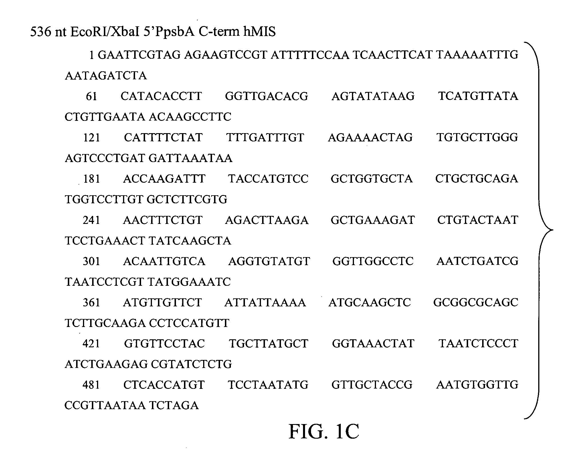
[0108] Based on the amino acid sequence of human MIS (GeneBank Accession # P03971) and our knowledge of chloroplast preferred codons, the carboxyl-terminal bioactive MIS DNA fragment was synthesized by BlueHeron Biotechnology, WA. The synthetic gene included an AUG start codon that is required for expression using chloroplast transformation technology (CTT), two 3′ translation stop codons, and a 5′ flanking EcoRI site and a 3′ flanking XbaI site for ease of subsequent cloning. See FIG. 1.

B. Construction of C-terminal MIS Plastid Transformation Expression Vectors:

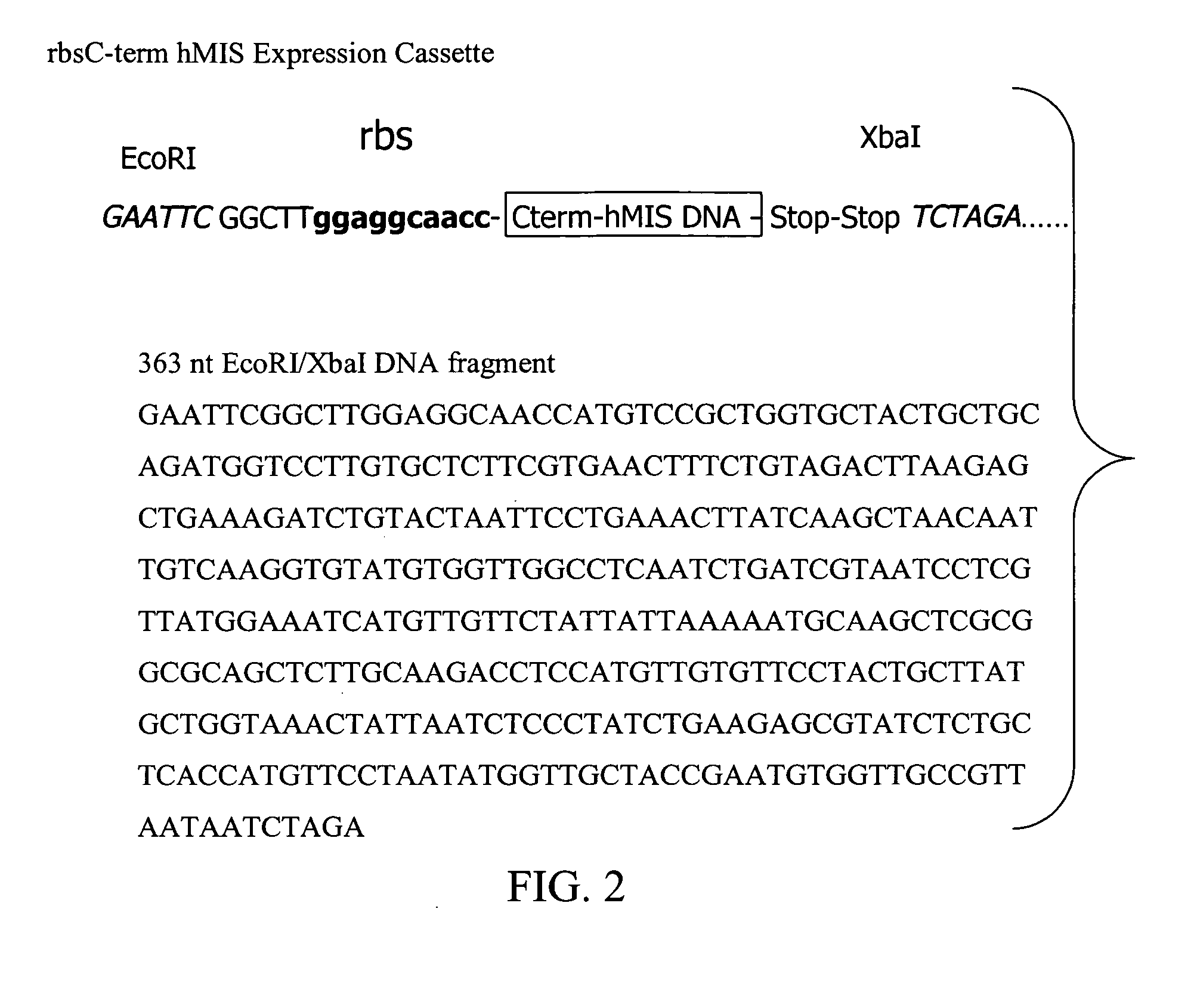
[0109] Two different 5′ regulatory sequences, a psbA gene promoter region and a Shine-Dalgamo ribosome binding site (rbs), were used to express the C-terminal MIS protein (C-term MIS). The Shine-Dalgarno sequence, 5′-AGGAGG-3′, is usually located 10 bp upstream of the AUG start codon and has been shown to be required for ribosom...

example 2

Plant Transformation

[0118] Tobacco (Nicotiana tabacum) plants grown aseptically by germination of seeds on MSO medium containing MS salts were used for transformation. As shown below, for each bombardment a whole leaf was placed abaxial side up on a Whatman filter paper on RMOP medium. DNA from the pLD MIS transformation expression plasmid clones was isolated using Qiagen Plasmid DNA Isolation Kit according to the manufacturer's recommendation (Qiagen, Inc). Gold (0.6 μm) microprojectiles were coated with greater than 900 ug / mL concentration of plasmid DNA containing the transgene. The bombardments and transformation were carried out with the biolistic device PDS 1000 / He (Bio-Rad) as described by Daniell H (1997) “Transformation and foreign gene expression in plants mediated by microprojectile bombardment”Meth. Mol. Biol. 62: 453-488. The bombarded tissue was placed on RMOP media for 36-48 hr prior to dissection into 5 mm squares which were then selected on 500 ug / ml spectinomycin ...

example 3

Bioassays

[0128] Once the MIS protein is purified from transplastomic plant tissue, the protein can be assayed for bioactivity using, for example, the standard organ culture bioassay for MIS (MacLaughlin, D. T., Hudson, P. L., Graciano, A. L., Kenneally, M. K., Ragin, R. C., Manganaro, T. F. & Donahoe, P. K. (1992) “Mullerian duct regression and antiproliferative bioactivities of Mullerian Inhibiting Substance reside in its carboxy-terminal domain”Endocrinology 131, 291-296) can be used to screen all plant generated samples for bioactivity and the results compared to purified MIS secreted from CHO cells (Cate, R. L., Mattaliano, R. J., Hession, C., Tizard, R., Farber, N. M., Cheung, A., Ninfa, E. G., Frey, A. Z., Gash, D. J., Chow, E. P., Fisher, R. A., Bertonis, J. M., Torres, G., Wallner, B. P., Ramachandran, K. L., Ragin, R. C., Manganaro, T. F., MacLaughlin, D. T. & Donahoe, P. K. (1986). Isolation of the bovine and human genes for Mullerian Inhibiting Substance and expression o...


PUM

PropertyMeasurementUnit
molecular weightaaaaaaaaaa
apparent molecular weightaaaaaaaaaa

Abstract

Bioactive TGF-β proteins are expressed in transgenic plastids. The TGF-β proteins are used for therapeutic and diagnostic purposes. In particular, Mullerian Inhibitor Substance (MIS), either full length or truncated proteins, are expressed in plastids and used for treating various cancers that contain MIS receptors such as ovarian cancer, breast cancer and prostate cancer.

Description

FIELD OF THE INVENTION [0001] The present inventions relates to the expression of bioactive TGF-β proteins in plastids. The TGF-β proteins are purified and used in diagnostic and therapeutic applications. In particular, Mullerian Inhibitor Substance (MIS) is made in plastids, purified and used to treat cancers that possess MIS receptors such as ovarian, breast and prostate cancers. BACKGROUND OF THE INVENTION [0002] Proteins of the TGF-.beta. family (TGF-β function to mediate many important embryogenic and immune functions including chemotaxis, production of extracellular matrix, regulation of cell growth and differentiation, and development and regulation of the immune system. Thus, these molecules could be used in a great variety of therapies if available in sufficient quantities. Epithelial ovarian cancers, for example, are the fifth most common malignancy in women. Each year approximately 26,600 new cases of epithelial ovarian cancer are diagnosed, of which 55% die of the diseas...

Claims


Application Information

Patent Timeline
Patent Type & Authority Applications(United States)
IPC IPC(8): A01H1/00A01H5/00C12N5/04C12N15/82A61K36/81
CPCC07K14/495C07K14/575C12N15/8214C12N15/8257
Inventor OISHI, KARENWILLIAMS, DAVID
Owner CHLOROGEN
Learn More