
Cationic liposome delivery of taxanes to angiogenic blood vessels

a technology of cationic liposomes and taxanes, which is applied in the field of liposome delivery, can solve the problems of not being able to fully appreciate the specificity of cationic liposomes for preferential targeting angiogenic endothelial cells, and not being able to clearly determine, so as to enhance wound healing, inhibit angiogenesis, and promote angiogenesis

Inactive Publication Date: 2005-12-08
RGT UNIV OF CALIFORNIA
View PDF25 Cites 4 Cited by
  • Summary
  • Abstract
  • Description
  • Claims
  • Application Information

AI Technical Summary

Benefits of technology

[0035] An advantage of the invention is that the cationic liposomes of the invention can be used to precisely deliver small amounts of toxic compounds to endothelial cells which cells are affected in a manner (e.g., killed) such that the blood vessel is destroyed or rendered inoperative such as by a blood clot and the nutrient supply to the surrounding tissues (such as tumor cells) is cut off thereby destroying the tissue (e.g., destroying a solid tumor).
[0036] Another advantage of the invention is that the cationic liposomes of the invention can be used to inhibit angiogenesis associated with malignant or benign tumors associated with on-going angiogenesis.
[0037] Yet another advantage of the invention is that the cationic liposomes can be used to provide for site directed delivery of compounds which promote angiogenesis and thereby enhance wound healing.
[0038] An important feature of the invention is that several classes of diseases and / or abnormalities are treated without directly treating the tissue involved in the abnormality e.g., by inhibiting angiogenesis the blood supply to a tumor is cut off and the tumor is killed without directly treating the tumor cells in any manner.
[0039] These and other objects, advantages and features of the present invention will become apparent to those skilled in the art upon reading the disclosure provided here in connection with the attached figures. BRIEF DESCRIPTION OF THE FIGURES
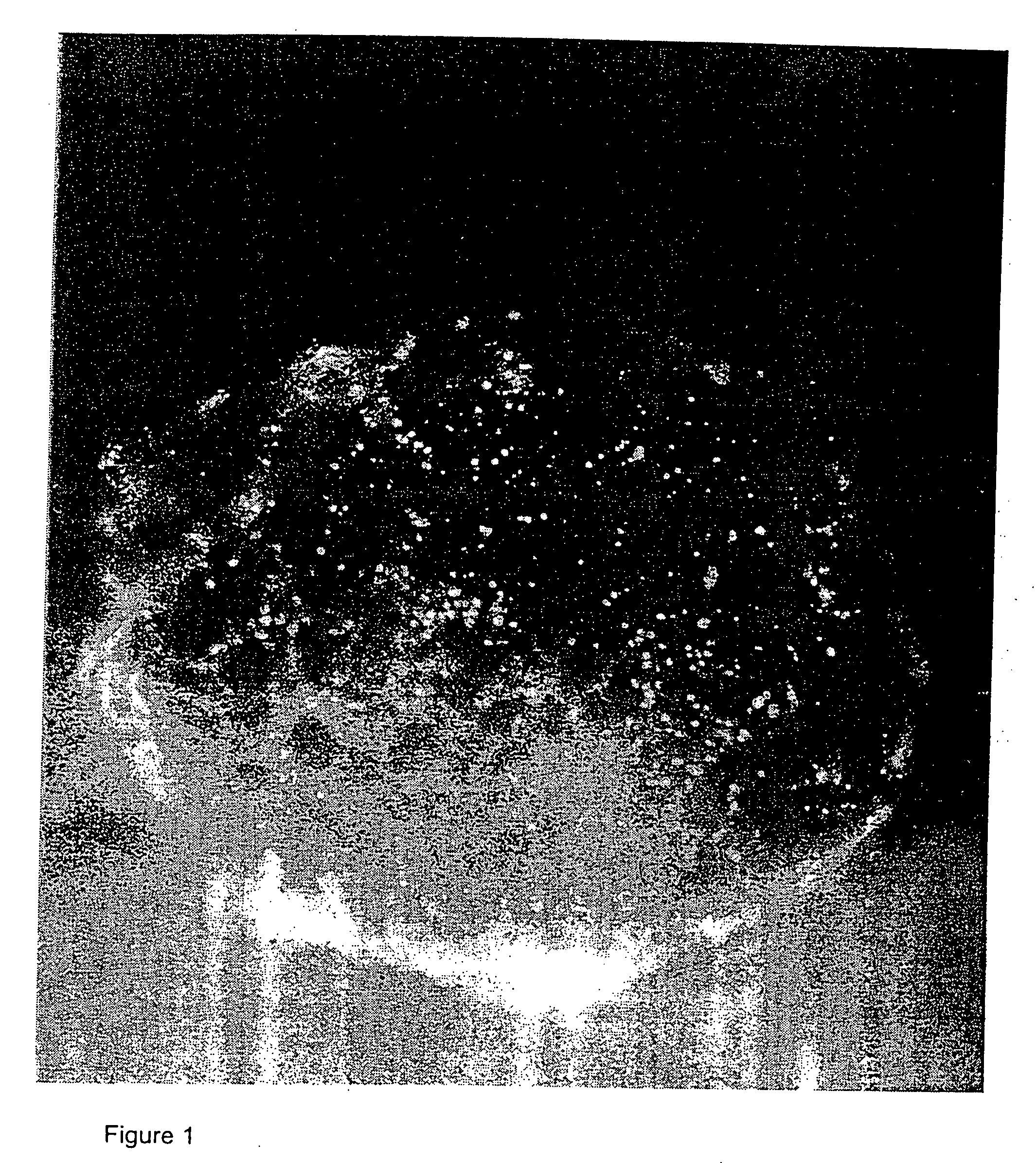
[0040]FIG. 1 is a fluorescence micrograph showing the uptake of red fluorescent CM-DiI-labeled DDAB:cholesterol-DNA complexes in angiogenic blood vessels of a follicle in a normal mouse ovary (Scale bar: 60 μm);

Problems solved by technology

However, within FIG. 1 it is not possible to clearly determine that the label is associated only with angiogenic endothelial cells or whether it is associated with all of the tissue within the ovary and follicle.
However, because so much of the label is associated with the angiogenic blood vessels in FIG. 4, it may not be possible to fully appreciate the specificity of cationic liposomes for preferentially targeting angiogenic endothelial cells.

Method used


Image

  • Cationic liposome delivery of taxanes to angiogenic blood vessels
  • Cationic liposome delivery of taxanes to angiogenic blood vessels
  • Cationic liposome delivery of taxanes to angiogenic blood vessels

Examples


example 1

Distribution of Cationic Liposomes in Normal Mice

[0150] Liposomes and / or the plasmid DNA were labeled and the cellular distribution of the labeled complexes at various times after intravenous injection were determined. The experiments were performed on pathogen-free mice (20-25 g body weight) of both sexes.

[0151] Cationic small unilamellar vesicle liposomes were prepared from the cationic lipid DDAB or DOTAP and the neutral lipid DOPE or cholesterol, labeled with Texas Red or the red fluorescent carbocyanine dye DiI or CM-DiI, and in some cases complexed to plasmid DNA containing a reporter gene such as luciferase or β-galactosidase. Endothelial cells were labeled using the fluorescent plant lectin fluorescein-Lycopersicon esculentum. Monocyte / macrophages were labeled by using fluorescent beads (Duke, 500 nm). Cell nuclei were labeled with DAPI, YO-PRO, or Hoechst 33342 dye.

[0152] Fluorescent liposomes or liposome-DNA complexes containing 10-60 μg of DNA in up to 300 μl were inje...

example 2

Uptake of DDAB:cholesterol Liposomes or Liposome-DNA Complexes in RIP-Tag5 Mice

[0162] The results of the experiments of Example 1 indicated that angiogenic blood vessels in ovarian follicles and corpora lutea avidly took up cationic liposomes and liposome-DNA complexes. Accordingly, an experiment was performed to determine whether endothelial cells of angiogenic blood vessels of tumors avidly take up cationic liposomes or liposome-DNA complexes.

[0163] The transgenic RIP-Tag5 model of tumors was used. see, Hanahan, (1985) Nature 315:115-22; and Hanahan and Folkman (1996) Cell 86:353-64. In this model, designated RIP-Tag, the oncogene from the SV-40 virus, large T antigen (Tag), is driven by a region of the rat insulin promoter (RIP). When inserted into the murine genome, this construct induces the expression of the T antigen specifically in β-cells of pancreatic islets.

[0164] One important attribute of this model is that various stages of tumor development, and therefore various s...

example 3

Uptake of DOTAP:cholesterol Liposomes-DNA Complexes in RIP-Tag2 Mice

[0168] Purpose: The fluorescence intensity of the liposome-DNA complexes had been increased by using Texas Red-DHPE in place of DiI; the method of preparing the pancreas of RIP-Tag2 mice for localizing sites of uptake of fluorescent cationic liposome-DNA complexes was improved; and the structure and function of the angiogenic blood vessels in pancreatic islet cell tumors in RIP-Tag2 mice had been studied. With these improvements experiments of the type described in Example 2 were carried out to determine how cationic liposomes and lipid-DNA complexes were taken up.

[0169] Methods: Cationic DOTAP:cholesterol small unilamellar vesicle liposomes, labeled with Texas Red-DHPE, were prepared. Liposome-DNA complexes were prepared at a total lipid:DNA ratio of 24:1 (nmoles / μg) in 5% glucose, using 60 μg of plasmid DNA in 300 μl. Complexes (300 μl) were injected into tail veins of unanesthetized transgenic RIP1-Tag2 C57BL / 6...


PUM

PropertyMeasurementUnit
diameteraaaaaaaaaa
diameteraaaaaaaaaa
diameteraaaaaaaaaa

Abstract

Angiogenic endothelial cells are selectively targeted with lipid / DNA complexes or cationic liposomes containing a substance which affects the targeted cells by inhibiting or promoting their growth. A site of angiogenesis can be precisely located by administering cationic liposomes containing a detectable label. The complexes may comprise nucleotide constructs which are comprised of promoters which are selectively and exclusively activated in the environment of an angiogenic endothelial cell.

Description

CROSS-REFERENCE TO RELATED APPLICATIONS [0001] This application is a continuation-in-part application of Ser. No. 09 / 127,177, filed Jul. 31, 1998, which is a continuation of Ser. No. 08 / 820,337, filed Mar. 12, 1997, now U.S. Pat. No. 5,837,283, both of which are incorporated herein by reference in their entirety and to which applications we claim priority under 35 U.S.C. §120.FIELD OF THE INVENTION [0002] The present invention is in the field of liposome delivery, and particularly of cationic liposome delivery of taxanes to blood vessels. BACKGROUND OF THE INVENTION [0003] The present invention can be applied to the treatment and diagnosis of a variety of different diseases and abnormalities. Although the present invention is not limited to such, it can be used in the treatment of cancer, wound healing, and a variety of chronic inflammatory diseases. In general, each is presently treated directly by physical means such as surgical removal of cancerous tissue, suturing of wounds and ...

Claims


Application Information

Patent Timeline
Patent Type & Authority Applications(United States)
IPC IPC(8): A61K9/127A61K31/337A61K47/48A61K49/00C12N15/88
CPCA61K9/1271A61K9/1272A61K31/335A61K31/337A61K49/0032A61K49/0041C12N15/88Y10S514/824A61K47/54A61K47/543A61P9/10
Inventor MCDONALD, DONALD M.MCLEAN, JOHN W.THURSTON, O. GAVIN
Owner RGT UNIV OF CALIFORNIA
Learn More