Anti-cancer antibodies with reduced complement fixation
a technology of anti-cancer antibodies and complement fixation, which is applied in the direction of peptides, drug compositions, and injected cells, can solve the problems of serious side effects of pain, and achieve the effects of reducing pain, complement fixation, and reducing pain
- Summary
- Abstract
- Description
- Claims
- Application Information
AI Technical Summary
Benefits of technology
Problems solved by technology
Method used
Image
Examples
example 1
Expression of an Anti-GD2 Antibody with a Mutation that Reduced Complement Fixation
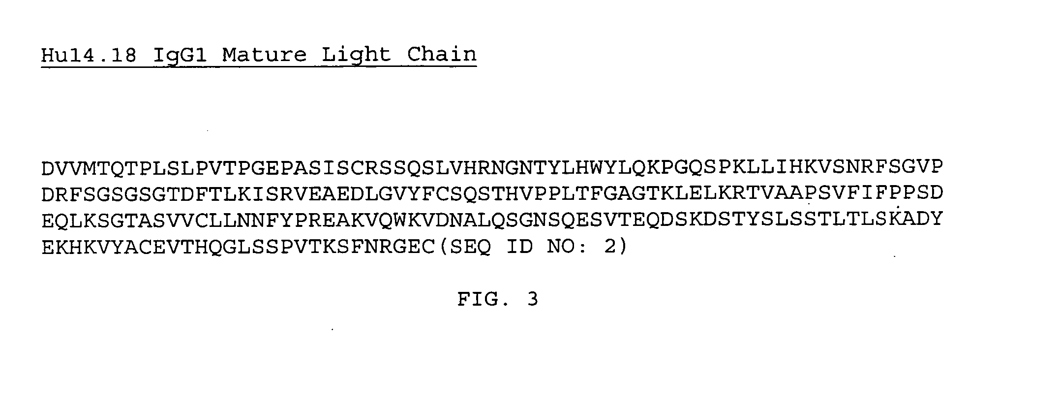
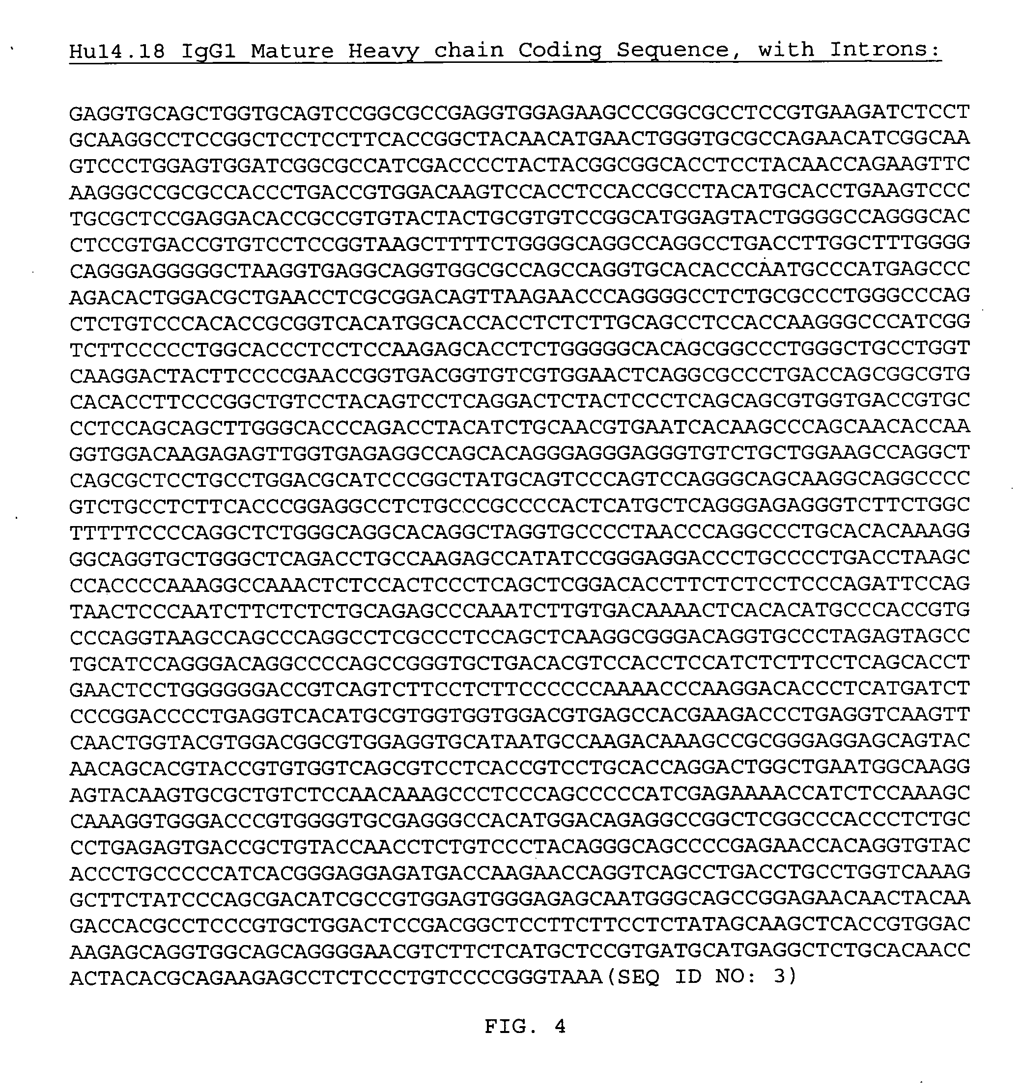
[0044] An expression plasmid that expresses the heavy and light chains of the human 14.18 anti-GD2 antibody with reduced complement fixation due to mutation was constructed as follows. The expression plasmid for the 14.18 anti-GD2 antibody was pdHL7-hu14.18. pdHL7 was derived from pdHL2 (Gillies et al., J. Immunol. Methods, [1989]; 125: 191-202), and uses the cytomegalovirus enhancer-promoter for the transcription of both the immunoglobulin light and heavy chain genes. The K322A mutation in the CH2 region was introduced by overlapping Polymerase Chain Reactions (PCR) using pdHL7-hu14.18 plasmid DNA as template. The sequence of the forward primer was 5′-TAC AAG TGC GCT GTC TCC AAC (SEQ ID NO:6), where the underlined GCT encodes the K322A substitution, and the sequence of the reverse primer was 5′-T GTT GGA GAC AGC GCA CTT GTA (SEQ ID NO:7), where the underlined AGC is the anticodon of the introduced a...
example 2
Transfection and Expression of the Anti-GD2 Antibody
[0045] Electroporation was used to introduce the DNA encoding the anti-GD2 antibody described above into a mouse myeloma NS / 0 cell line or the YB2 / 0 cell line. To perform electroporation, cells were grown in Dulbecco's modified Eagle's medium supplemented with 10% heat-inactivated fetal bovine serum, 2 mM glutamine and penicillin / streptomycin. About 5×10⁶ cells were washed once with PBS and resuspended in 0.5 ml PBS. 10 μg of linearized plasmid DNA encoding the modified anti-GD2 antibody of Example 1 was then incubated with the cells in a Gene Pulser Cuvette (0.4 cm electrode gap, BioRad) on ice for 10 min. Electroporation was performed using a Gene Pulser (BioRad, Hercules, Calif.) with settings at 0.25 V and 500 μF. Cells were allowed to recover for 10 min on ice, after which they were resuspended in growth medium and plated onto two 96 well plates.
[0046] Stably transfected clones were selected by their growth in the presence o...
example 3
Biochemical Analysis of the Anti-GD2 Antibody
[0047] Routine SDS-PAGE characterization was used to assess the integrity of the modified antibodies. The modified anti-GD2 antibodies were captured on Protein A Sepharose beads (Repligen, Needham, Mass.) from the tissue culture medium into which they were secreted, and were eluted by boiling in protein sample buffer, with or without a reducing agent such as β-mercaptoethanol. The samples were separated by SDS-PAGE and the protein bands were visualized by Coomassie staining. Results from SDS-PAGE showed that the modified anti-GD2 antibody proteins analyzed were present substantially as a single band, indicating that there was not any significant amount of degradation.
PUM
| Property | Measurement | Unit |
|---|---|---|
| Fraction | aaaaa | aaaaa |
| Frequency | aaaaa | aaaaa |
| Frequency | aaaaa | aaaaa |
Abstract
Description
Claims
Application Information
 - R&D
- Intellectual Property
- Life Sciences
- Materials
- Tech Scout
- Unparalleled Data Quality
- Higher Quality Content
- 60% Fewer Hallucinations
Browse by: Latest US Patents, China's latest patents, Technical Efficacy Thesaurus, Application Domain, Technology Topic, Popular Technical Reports.
© 2025 PatSnap. All rights reserved.Legal|Privacy policy|Modern Slavery Act Transparency Statement|Sitemap|About US| Contact US: help@patsnap.com


