
Method of regenerating blood vessels

Inactive Publication Date: 2003-09-25
ASAHI KASEI MEDICAL CO LTD
View PDF8 Cites 13 Cited by
  • Summary
  • Abstract
  • Description
  • Claims
  • Application Information

AI Technical Summary

Benefits of technology

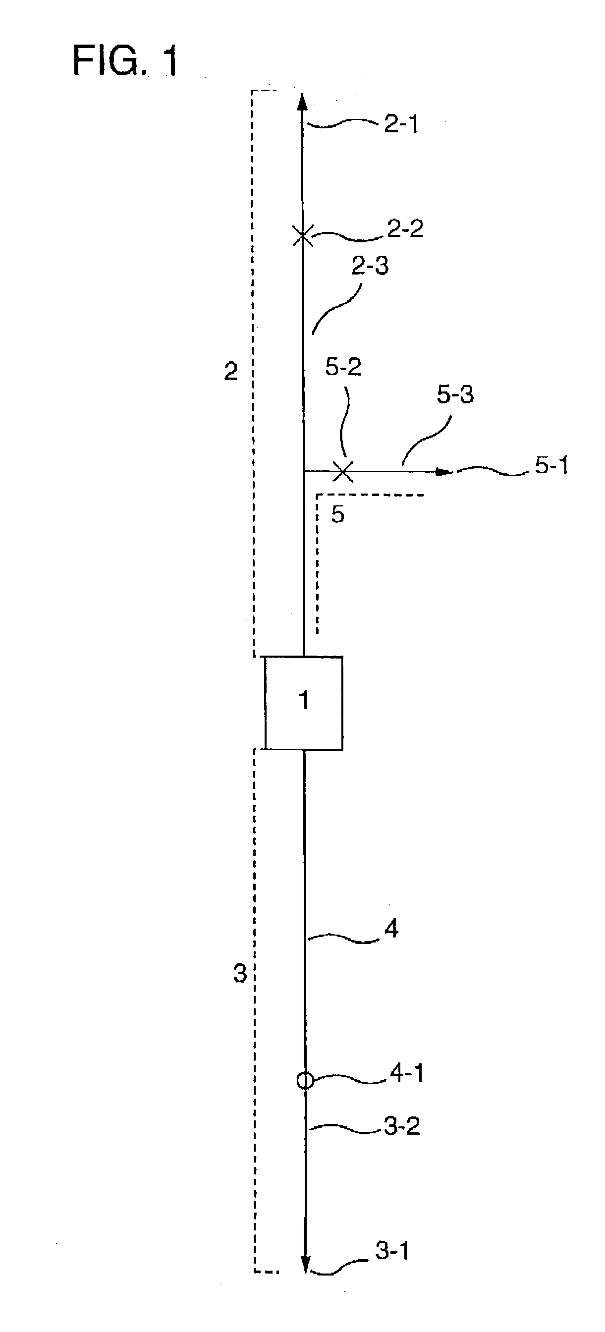
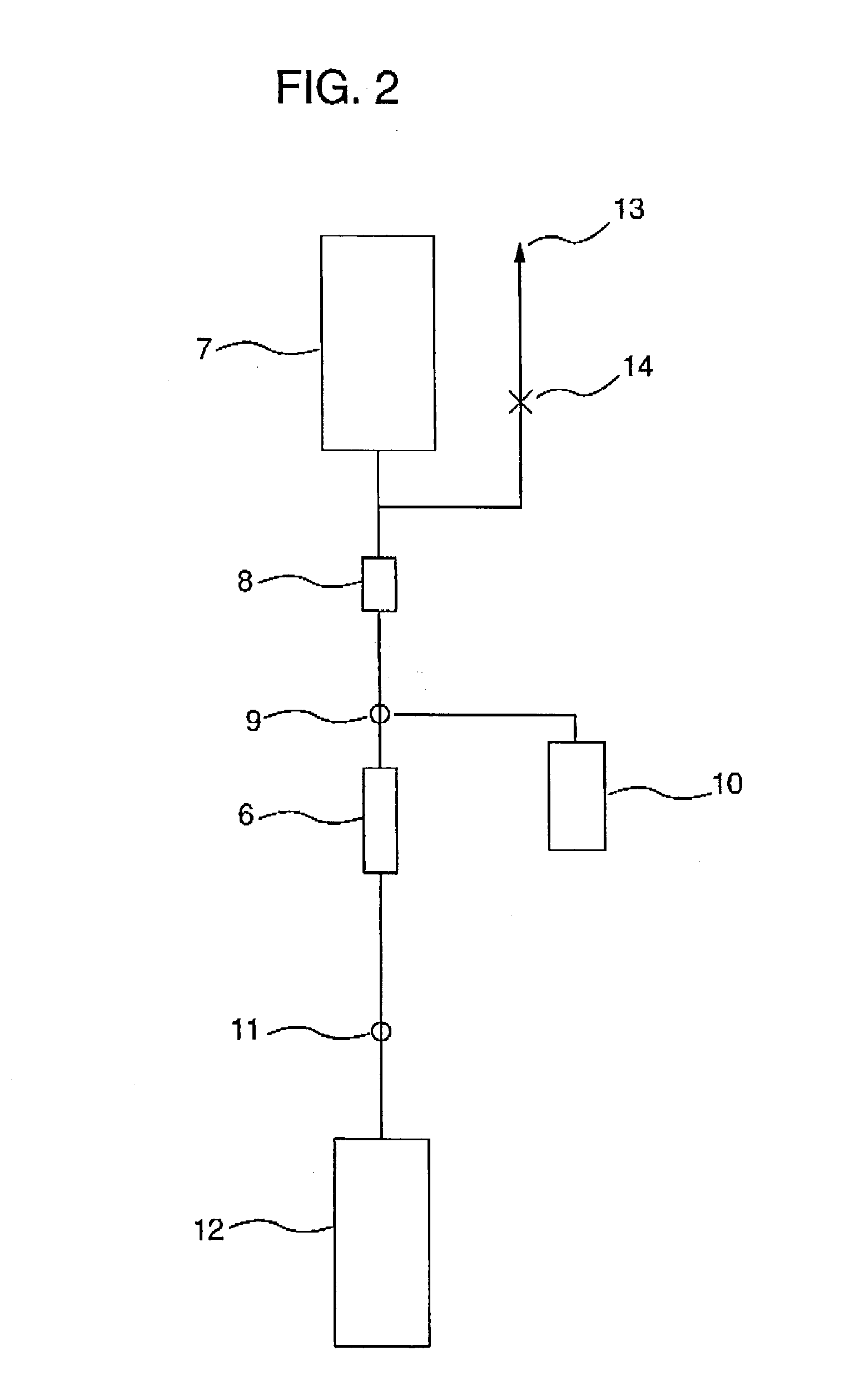
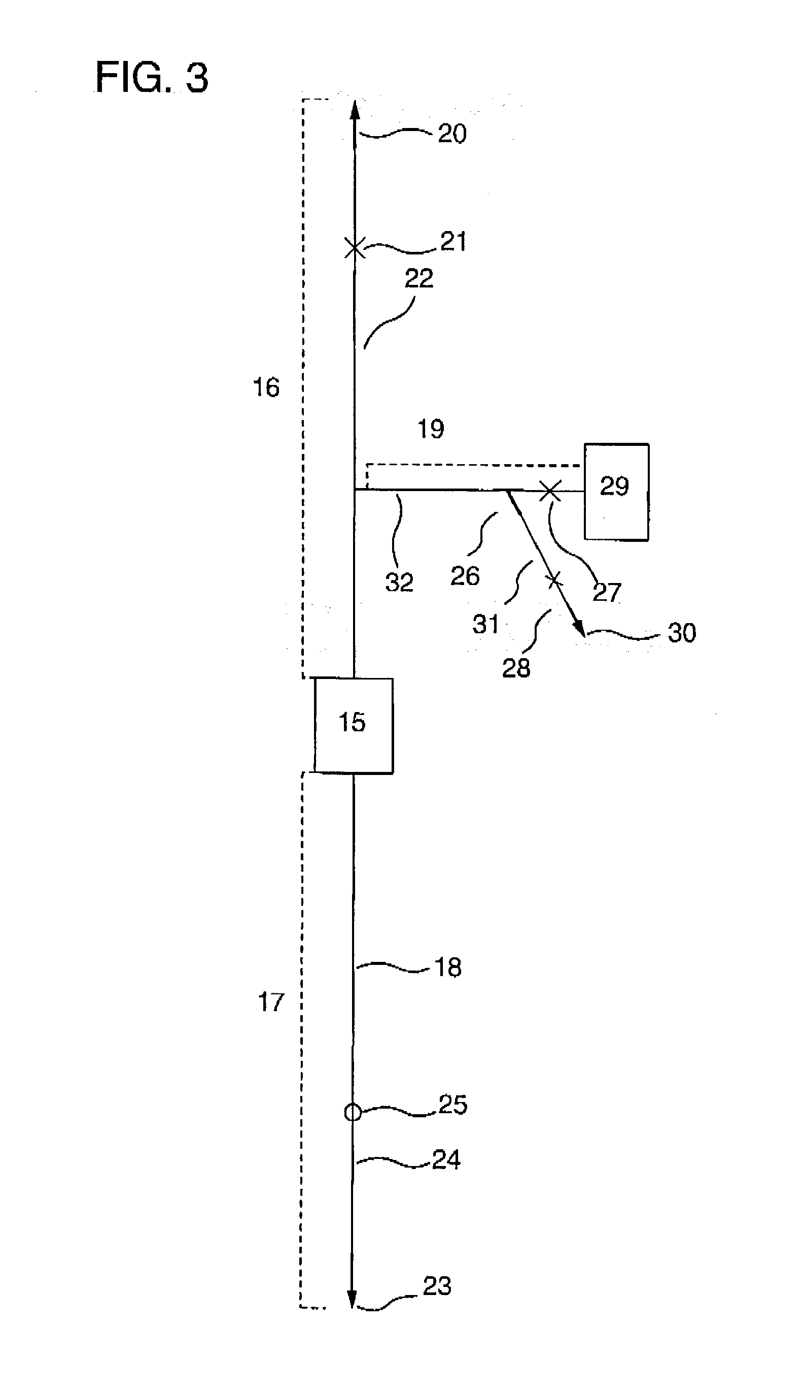
[0059] In the present invention, the cell-capturing means capable of capturing at least cells to be recovered and substantially permitting passage therethrough of cells to be removed, may comprise a container having a liquid inlet and a liquid outlet which is packed with a material capable of capturing the cells to be recovered and substantially permitting passage therethrough of the cells to be removed, and a molded container having a cell-capturing surface on its inner surface. In the present invention, "cell-capturing means" may be called "cell separator". The material capable of capturing the cells to be recovered and substantially permitting passage therethrough of the cells to be removed may be any conventional cell-capturing material so long as it can selectively capture the cells to be recovered. The following materials, for example, are preferable because of their excellent moldability, sterilizability, and low cytotoxicity: synthetic polymers such as polyethylenes, polypropylenes, polystyrenes, acrylic resins, nylons, polyesters, polycarbonates, polyacrylamides, polyurethanes, etc; natural polymers such as agarose, cellulose, cellulose acetate, chitin, chitosan, alginates, etc.; inorganic materials such as hydroxyapatite, glass, alumina, titania, etc.; and metals such as stainless steel, titanium, aluminum, etc.
[0084] As a method for introducing the cell-containing fluid referred to in the present specification into the cell-capturing means, there may be adopted either a method of connecting a bag or bottle containing the cell-containing fluid through a tube, and then introducing the fluid, for example, by utilizing its fall, a roller pump, causing a flow of the fluid by squeezing the bag, or by a method of connecting a syringe containing the cell-containing fluid, and introducing the fluid by pushing the piston of the syringe by hand or using a device such as a syringe pump. The pushing by hand is characterized by its simplicity, and the use of the device is characterized in that the control of the flow rate of the recovering liquid in its introduction is easy. Therefore, a suitable method is selected depending on the purpose.
[0093] The "transvascular transplantation" in the present invention is the transplantation of the recovered vascular endothelial precursor cells or the attaching cells obtained by further cultivating them via blood vessel because of easy introduction to the patient. The blood vessel to which the cells are introduced through a needle Or a catheter is preferably the one in lower extremities or superior limb. The total amount of vascular endothelial precursor cells via blood vessel is preferably 2-10 times as much as those transplanted directly, since the efficiency to reach to the ischemic region is lower than that of direct transplantation.
[0108] The present invention still further provides a liquid containing cells to be recovered and substantially no cells to be removed which is obtained by a cell separation method comprising steps of introducing a cell-containing fluid containing cells to be recovered and cells to be removed, into a cell-capturing means capable of substantially capturing said cells to be recovered and substantially permitting passage there-through of said cells to be removed. The resulting fluid containing the cells to be removed is taken out of the cell-capturing means, and then a liquid with a viscosity of not more than 500 mPa.s and not less than 5 mPa.s is introduced into the cell-capturing means to recover the cells which have been captured by the cell-capturing means. When the separation method of the present invention is applied to a suspension containing cells to be recovered and cells to be removed, it becomes possible to efficiently provide a suspension substantially comprising the cells to be recovered.

Problems solved by technology

The term "cells to be removed" means cells unnecessary for the above purpose or cells which should be positively removed because they are, for example, pathogenic cells, so that contamination by them of cells to be recovered causes a problem.

Method used


Image

  • Method of regenerating blood vessels
  • Method of regenerating blood vessels
  • Method of regenerating blood vessels

Examples


example 1

[0115] This working example shows an example of cell separation in the case where a cell-containing fluid was cord blood, cells to be recovered are a mononuclear cell fraction containing hematopoietic stem cells, and cells to be removed are erythrocytes and platelets.

[0116] {circle over (1)} Cell Separator

[0117] A polycarbonate container with outside dimensions (length.times.width.times.thickness) of 41.times.41.times.18 mm having a liquid outlet and a liquid inlet on the diagonal was packed with 12 polyester nonwoven fabrics with an average fiber diameter of 2.3 .mu.m on the inlet side and 25 polyester nonwoven fabrics with an average fiber diameter of 12 .mu.m on the outlet side. The packing density was 0.24 g / cm.sup.3, the effective filtration area 9 cm.sup.2, and the effective filtration length 12.4 mm. In order to impart platelet permeability to the resulting filter, coating with a hydrophilic polymer was carried out. In detail, a 1% ethanolic solution of a hydroxyethyl methacr...

example 2

[0139] This working example shows an example of cell separation in the case where a cell-containing fluid was peripheral blood, cells to be recovered are leukocytes, and cells to be removed are erythrocytes and platelets.

[0140] {circle over (1)} Cell Separator

[0141] A polycarbonate container with outside dimensions (length.times.width.times.thickness) of 41.times.41.times.18 mm having a liquid outlet and a liquid inlet on the diagonal was packed with 25 polyester nonwoven fabrics with an average fiber diameter of 12 .mu.m on the inlet side and 12 polyester nonwoven fabrics with an average fiber diameter of 2.3 .mu.m on the outlet side. The packing density was 0.24 g / cm.sup.3, the effective filtration area 9 cm.sup.2, and the effective filtration length 12.4 mm. In order to impart platelet permeability to the resulting filter, coating with a hydrophilic polymer was carried out. A 1% ethanolic solution of a hydroxyethyl methacrylate dimethylaminoethyl methacrylate copolymer (molar rat...

example 3

[0148] This working example shows an example of cell separation in the case where a cell-containing fluid was cord blood, cells to be recovered are hematopoietic stem cells (CD34-positive cells), and cells to be removed are erythrocytes and platelets.

[0149] {circle over (1)} Cell Separator

[0150] A polycarbonate container with outside dimensions (length.times.width.times.thickness) of 41.times.41.times.18 mm having a liquid outlet and a liquid inlet on the diagonal was packed with 12 polyester nonwoven fabrics with an average fiber diameter of 12 .mu.m on the inlet side and 25 polystyrene nonwoven fabrics with an average fiber diameter of 2.3 .mu.m having anti-human CD34 monoclonal mouse antibody (clone name: Immu133, available from Coulter Corp.; hereinafter abbreviated as "CD34 antibody") immobilized thereon, on the outlet side. The packing density of the resulting filter was 0.2 g / cm.sup.3. The immobilization of the anti-human CD34 monoclonal mouse antibody on-the polystyrene was ...


PUM

PropertyMeasurementUnit
viscosityaaaaaaaaaa
viscosityaaaaaaaaaa
diameteraaaaaaaaaa

Abstract

A method for regenerating a blood vessel comprising introducing a cell-containing fluid containing vascular endothelial precursor cells and cells to be removed into a cell separator which allows at least the cells to be removed to substantially pass through but substantially captures the vascular endothelial precursor cells; recovering the vascular endothelial precursor cells once captured on said cell separator by introducing a fluid into the said cell separator; and using the recovered vascular endothelial precursor cells for regenerating a blood vessel.

Description

[0001] This application is a continuation-in-part of U.S. patent application Ser. No. 09 / 871,645 filed Jun. 4, 2001, which is a divisional application of U.S. patent application Ser. No. 09 / 341,879 filed Jul. 19, 1999, the entire contents of both of which are hereby incorporated by reference.[0002] (1) Field of the Invention[0003] This invention relates to a method for separating and recovering only necessary cells from a fluid containing a mixture of various cells. The cells thus obtained can be used in providing therapy for various diseases, such as hematopoietic stem cell transplantation, and in fundamental sciences such as immunology and cell biology.[0004] (2) Description of the Related Art[0005] Japanese patent JP-A-54-119012 discloses a technique for recovering lymphocytes by capturing leukocytes on a filter from a body fluid such as blood containing leukocytes (granulocytes, monocytes and lymphocytes) and erythrocytes.[0006] In the case of hematopoietic stem cell transplanta...

Claims


Application Information

Patent Timeline
Patent Type & Authority Applications(United States)
IPC IPC(8): A61K35/12C12N5/00C12N5/074
CPCA61K35/12C12N5/0692C12N5/0087
Inventor MUROHARA, TOYOAKIAOKI, MIKASUMITA, MASAYATERASHIMA, SHUJIYASUTAKE, MIKITOMO
Owner ASAHI KASEI MEDICAL CO LTD
Learn More