Extracellular matrix for treating pelvic floor disorders and skeletal muscle degeneration
a technology of extracellular matrix and pelvic floor, which is applied in the field of extracellular matrix for treating pelvic floor disorders and skeletal muscle degeneration, and can solve problems such as poor cell survival and increased connective tissu
- Summary
- Abstract
- Description
- Claims
- Application Information
AI Technical Summary
Benefits of technology
Problems solved by technology
Method used
Image
Examples
example 1
Injectable Skeletal Muscle Matrix
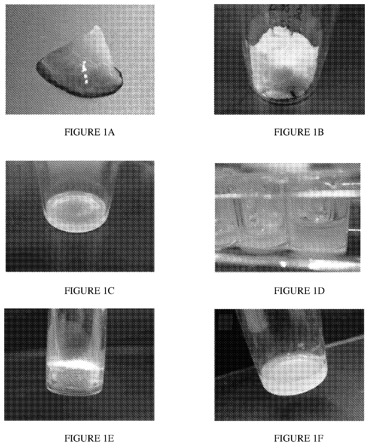
[0105]Skeletal muscle matrix material was derived through decellularization of porcine skeletal muscle tissue (FIG. 1A). Fat and connective tissue was removed, and the skeletal muscle was cut into ˜1 cm3 pieces, rinsed with deioninized water and stirred in 1% (wt / vol) solution of SDS in PBS for 4-5 days. The decellularized muscle was then stirred overnight in deionized water, and agitated rinses under running deionized water were performed to remove residual SDS. In addition to confirmation with lack of nuclei on H&E stained sections (not shown), the DNA content of the material was measured as 26.14±1.67 ng of DNA / mg of dry weight ECM, which confirmed decellularization. The matrix was then lyophilized (FIG. 1B) and milled into a fine particulate. At this stage the material can be hydrated and utilized for in vivo injection or it can be enzymatically digested to form a liquid (FIG. 1C). At this stage, the liquid skeletal muscle matrix can be diluted a...
example 2
Mitogenic Assay
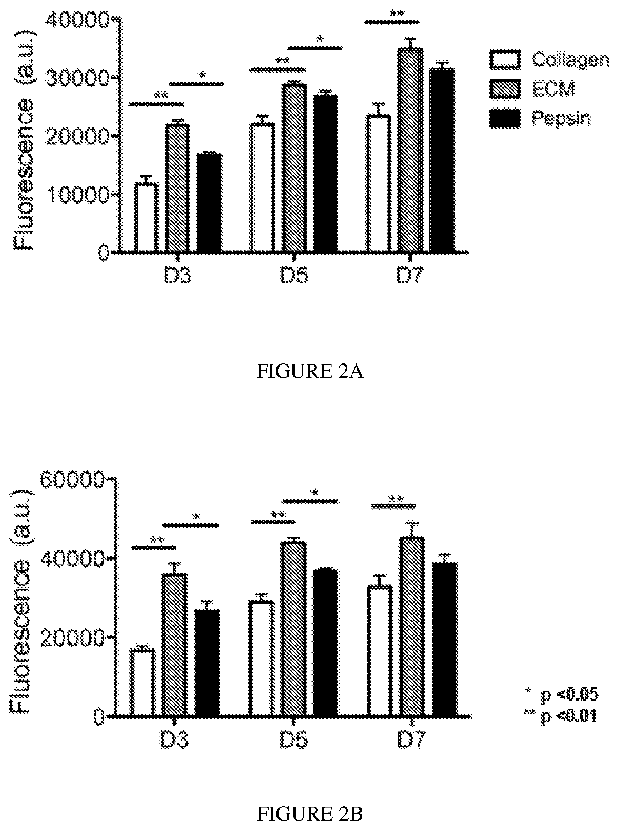
[0106]Degradation products of decellularized ECMs have been previously shown to have mitogenic activity. It was examined whether the degradation products of the skeletal muscle matrix hydrogel had a mitogenic effect on cells in vitro. Proliferation of smooth muscle cells and skeletal myoblasts following exposure to either enzymatically degraded skeletal muscle ECM or collagen was assessed. Pepsin was also included as a control, as pepsin was utilized to digest the matrix material. A Picogreen assay was used to determine double stranded DNA content at days 3, 5, and 7 in culture to quantify cell proliferation. It was found that both smooth muscle cells (FIG. 2A) and myoblasts (FIG. 2B), when cultured in media containing degraded skeletal muscle matrix, had a higher rate of proliferation compared to cells cultured in media containing the same concentration of collagen. The increase in cell number was significantly greater at all time points (p<0.01). At day 3, there was...
example 3
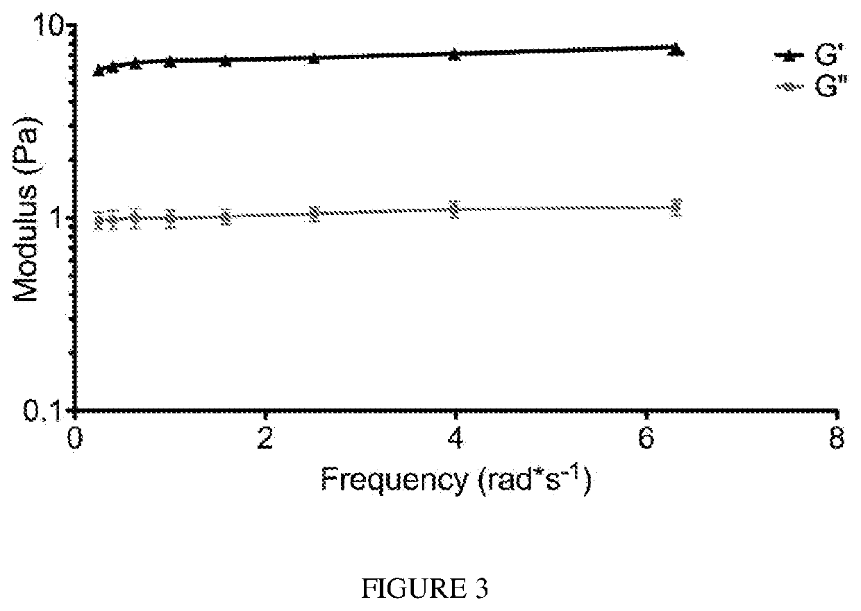
[0107]A gel form of the matrix was initially made in vitro by bringing the material (6 mg / mL) to a physiological pH and incubating the material at 37° C. After gelation, the material was tested for rheological properties where it was determined that the material had a storage modulus (G′) of 6.5±0.5 Pa. A representative trace of rheological data is shown in FIG. 3. The ability of the liquid skeletal muscle matrix to form a gel in situ were then assessed by injecting the material into a healthy rat hindlimb. For all in vivo studies, liquid skeletal muscle matrix, which had been biotinylated and re-lyophilized for storage at −80 C were utilized. Prior to injection, the material was resuspended in sterile water alone. The skeletal muscle matrix was then loaded into a syringe and injected intramuscularly into a rat hindlimb (FIG. 4A). To determine whether the skeletal muscle matrix would assemble and form a scaffold, the injection region was excised after 20...
PUM
| Property | Measurement | Unit |
|---|---|---|
| temperature | aaaaa | aaaaa |
| temperature | aaaaa | aaaaa |
| concentration | aaaaa | aaaaa |
Abstract
Description
Claims
Application Information
 - R&D
- Intellectual Property
- Life Sciences
- Materials
- Tech Scout
- Unparalleled Data Quality
- Higher Quality Content
- 60% Fewer Hallucinations
Browse by: Latest US Patents, China's latest patents, Technical Efficacy Thesaurus, Application Domain, Technology Topic, Popular Technical Reports.
© 2025 PatSnap. All rights reserved.Legal|Privacy policy|Modern Slavery Act Transparency Statement|Sitemap|About US| Contact US: help@patsnap.com


