Method for separating and culturing mesenchymal stem cells from Wharton's jelly tissue of umbilical cord
a mesenchymal stem cell and umbilical cord technology, applied in the field of stable method for separating and extracting mesenchymal stem cells from umbilical cord jelly tissue, can solve the problems of quality and quantity of cord mesenchymal stem cells, and achieve the effects of ensuring the viability of stem cells, reducing interference from red blood cells, and improving cell purity
- Summary
- Abstract
- Description
- Claims
- Application Information
AI Technical Summary
Benefits of technology
Problems solved by technology
Method used
Image
Examples
example 1
Screening of the Components of Serum-Free Medium for Mesenchymal Stem Cells
[0088](1) Screening of the Content of Serum Substitute
[0089]Medium to be tested: 0.1 parts by volume of β-mercapto, 10 ng / ml recombinant human basic fibroblast growth factor (b-FGF, Peprotech), 1 part by volume of aqueous solution of non-essential amino acids (Catalog No. 11140, Gibco), 1, 2, 5, 8, 10, 12, 15, or 20 parts by volume of Knockout FBS serum substitute (Catalog No. 10828-028, Gibco), and 89 parts by volume of a-MEM.
[0090]In a biosafety cabinet, the third-passage hUC-MSCs separated from Wharton's jelly tissue of umbilical cord from a healthy neoborn by natural delivery were inoculated into a T75 cell culture flask at a density of 2×10⁴ cells / cm2, and 12-15 ml of medium commercially available were added as well to culture the cells. The cells were cultured till cells were observed to have completely adhered to walls of the flask, and the medium was replaced with 15 ml of medium to be tested. The gro...
example 2
Red Blood Cell Lysis Buffer Assisted Method of Extracting Umbilical Cord Mesenchymal Stem Cells
[0098]Collecting and Transporting the Sample:
[0099]A umbilical cord tissue sample from a newborn by cesarean section delivery was collected aseptically, put into the preservation and transportion solution for umbilical cord comprising penicillin sodium, streptomycin sulfate, gentamicin and amphotericin B (the final concentration of each of penicillin sodium, streptomycin sulfate and gentamicin in D-Hank's was 150 U / ml respectively, while that of amphotericin B was 300 U / ml.), then transported in ice to a clean GMP cell laboratory within 2 hours.
[0100]Washing and Sterilizing the Sample:
[0101]In a biosafety cabinet, the fresh umbilical cord sample was placed in a 50 ml aseptic centrifuge tube, washed 2 times with 75% aqueous ethanol, then washed 3 times with sterile saline.
[0102]Pre-Processing the Umbilical Cord Sample:
[0103]The umbilical cord was cut into segments each of about 2-3 cm in le...
example 3
Red Blood Cell Lysis Buffer Assisted Method of Extracting Umbilical Cord Mesenchymal Stem Cells
[0110]The red blood cell lysis buffer used in this Example comprised 10 g / L NH4Cl and 0.1 mM Na2-EDTA, pH 7.2-7.4.
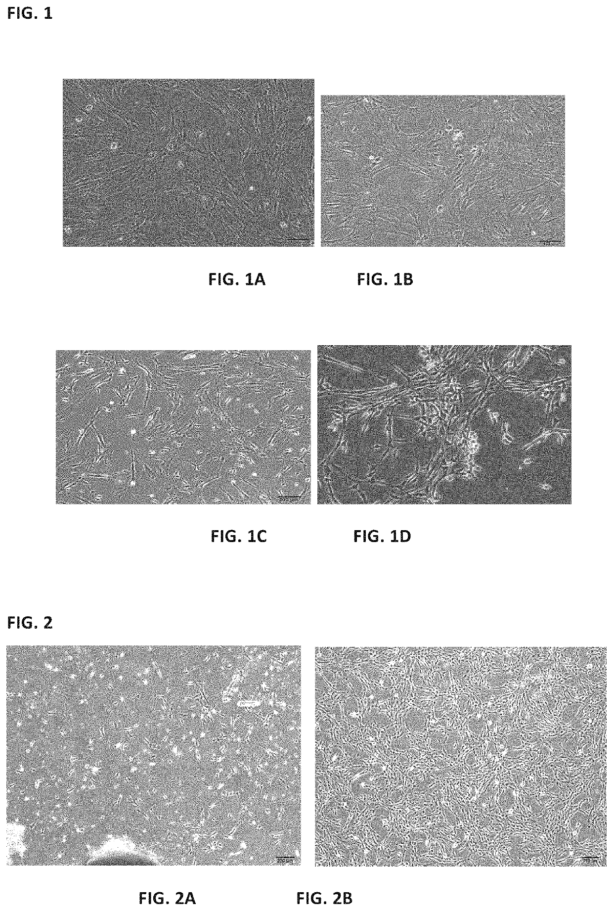
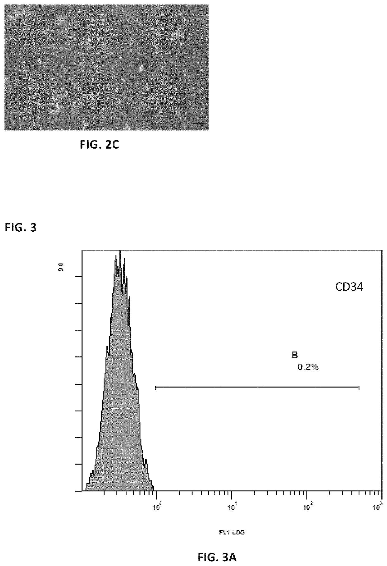
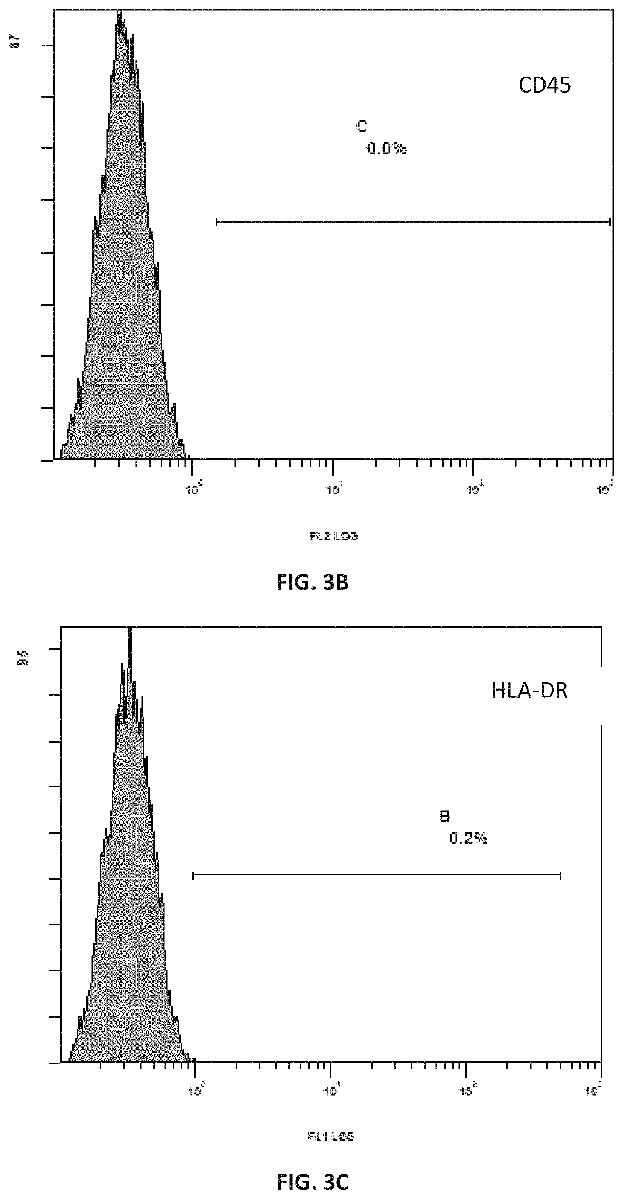
[0111]The process was carried out with reference to the method described in Example 2 above. The mesenchymal stem cells grew out of the bottom of the tissue blocks on the 5th day of culture, and formed cell-clusters in whorls around the 7th day of culture. After the removal of the tissue blocks and replacement with fresh medium, 60% confluence was reached around 12th day, and the cells were passaged after trypsin digestion. The purity of the cells after three passages was greater than 99% while the viability was more than 95%.
PUM
| Property | Measurement | Unit |
|---|---|---|
| concentration | aaaaa | aaaaa |
| concentration | aaaaa | aaaaa |
| length | aaaaa | aaaaa |
Abstract
Description
Claims
Application Information
 - R&D
- Intellectual Property
- Life Sciences
- Materials
- Tech Scout
- Unparalleled Data Quality
- Higher Quality Content
- 60% Fewer Hallucinations
Browse by: Latest US Patents, China's latest patents, Technical Efficacy Thesaurus, Application Domain, Technology Topic, Popular Technical Reports.
© 2025 PatSnap. All rights reserved.Legal|Privacy policy|Modern Slavery Act Transparency Statement|Sitemap|About US| Contact US: help@patsnap.com


