
Methods and devices for diagnosis of particles in biological fluids

a biological fluid and particle technology, applied in the field of methods and devices for diagnosing particle can solve the problems of limited sensitivity, inability to meet the needs of different operators, inability to diagnose particles in biological fluids, etc., and achieve the effects of reducing the unacceptably high misdiagnosis rate of crystal species, and reducing the misdiagnosis ra

Active Publication Date: 2018-08-28
CASE WESTERN RESERVE UNIV
View PDF11 Cites 0 Cited by
  • Summary
  • Abstract
  • Description
  • Claims
  • Application Information

AI Technical Summary

Benefits of technology

[0014]In view of the above, another problem of the present invention is to provide a method that distinguishes between monosodium urate monohydrate and calcium pyrophosphate dihydrate compounds, in the form of crystals, that accurately distinguish between the compounds that cause gout and pseudogout diseases respectively.
[0017]A further object of the present invention is to provide an affordable, automated and portable technology that can be utilized in a primary care setting for reducing the unacceptably high misdiagnosis rate of crystal species in joint spaces. A lower misdiagnosis rate would impact patients' quality of life as it translates to less suffering, less deterioration of joints and avoidance of incorrect treatments, introducing cost savings.
[0019]Yet another object of the present invention is to provide a method that is affordable, automated, and readily available to primary care facilities that can accurately identify pathologic crystals in biological fluids, namely synovial aspirates of patients with gout-like symptoms or urethral or kidney stones in urine samples.
[0020]Yet another object of the present invention is to provide a method for determining whether certain compounds, in particular crystals, are present in a sample, in particular a sample of synovial fluid, including the step of performing a digestion step of the fluid which has the effect of releasing, when present, marker compounds or crystals, in particular from an organic material or reducing the viscosity of the fluid.

Problems solved by technology

These crystals cause inflammation, pain and destruction in the joint.
However, the treatment of gout and pseudogout and different because the crystals accumulated are not the same.
Microscopic polarized imaging (PLM), which is presently the clinical standard of identifying pathologic crystals in synovial aspirates of patients with gouty symptoms, has limited sensitivity, and is prone to the subjectivity of the different operators.
Currently, false negative rate in gout is 30%, and false positives happen up to 20% of the time has been reported in the literature, lead to inappropriate long-term treatment and delay the treatment of the actual cause.
This strategy necessitates the utilization of research-grade Raman instruments with premium signal collection capability and, thus, limits the translation of the method to the clinical applications.
First, the stone is not diagnosed until it has formed enough mass to be identified by the imaging studies such as CT scans.
Second, the composition of the stone is not identified until and when the stone is removed or passed spontaneously.
Third, in majority of modern day surgery, powerful energy sources are used to powder fragments of the stones so the patient could pass them and therefore the fragments of the stone may not be obtained for future analysis.
Fourth, there are no available technologies to monitor the status of those patients with high risk of redeveloping stones, or to assess their responsiveness to treatment.
Most physicians do not monitor high risk patients due to the absence of a practical diagnostic method that can be executed at the point of care.
Imaging modalities such as CT, X-rays and ultrasonography do not provide specific information on the type and composition of stones.
Available techniques for stone compositional analysis (e.g. wet chemical analysis, infrared spectroscopy, x-ray diffraction, and scanning electron microscopy) are performed by specialty labs using bulky and expensive equipment.
Further, there are indirect costs associated with shipping and handling samples and coordinating the test results post hoc.

Method used


Image

  • Methods and devices for diagnosis of particles in biological fluids
  • Methods and devices for diagnosis of particles in biological fluids
  • Methods and devices for diagnosis of particles in biological fluids

Examples


examples

[0053]Set A

[0054]Sample Collection and Preparation

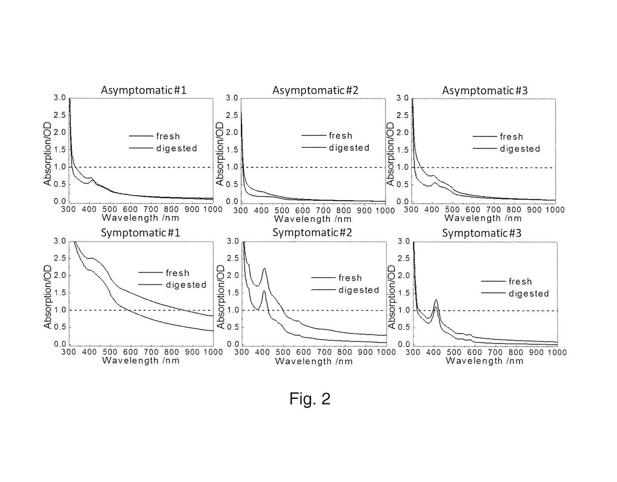
[0055]Synovial aspirates were collected from seven patients presenting “gout-like symptoms” and three asymptomatic synovial aspirates obtained from Anatomy Gifts Registry (Hanover, Md.) where the donors did not have any known joint disease history. Symptomatic sample collection was conducted under the approvals of Institutional Review Boards of institutions where synovial samples were collected (Metro Health Hospital, Cleveland, Ohio and Henry Ford Hospital, Detroit, Mich.). The patients presented to the clinic with gout-like symptoms and aspirates were collected as part of the normal diagnostic procedure. Symptomatic samples had aggregates within the fluid whereas the asymptomatic samples were clear in appearance. The synovial fluid samples were frozen following collection and kept frozen during shipment and long-term storage at −20° C. The presence of MSU crystals were confirmed by compensated polarized imaging as known in the art....


PUM

PropertyMeasurementUnit
excitation wavelengthaaaaaaaaaa
poweraaaaaaaaaa
concentrationaaaaaaaaaa

Abstract

Methods for determining whether certain compounds, in particular crystals, are present in a sample of a biological fluid that indicates an individual has a particular disease or condition, such as but not limited to gout, pseudogout or urinary tract stones. In some embodiments, the methods include the steps of digestion and filtration of a sample of synovial fluid in order to isolate, if present, monosodium urate monohydrate (MSU), calcium pyrophosphate dihydrate (CPPD), or calcium phosphate crystals from the sample, wherein the filtrate is analyzed with a Raman device to ascertain the presence and type of the crystals. Devices for performing steps of the method are disclosed.

Description

[0001]This invention was made with government support under contract number R01AR057812 awarded by the National Institutes of Health. The government has certain rights in the invention.FIELD OF THE INVENTION[0002]The present invention relates to methods for determining whether certain compounds, in particular crystals, are present in a sample of a biological fluid that indicates an individual has a particular disease or condition, such as but not limited to gout, pseudogout or urinary tract stones. In some embodiments, the methods include the steps of digestion and filtration of a sample of synovial fluid in order to isolate, if present, monosodium urate monohydrate (MSU), calcium pyrophosphate dehydrate (CPPD), or calcium phosphate crystals from the sample, wherein the filtrate is analyzed with a Raman device to ascertain the presence and type of the crystals. Devices for performing steps of the method are disclosed.BACKGROUND OF THE INVENTION[0003]Various biological fluids include...

Claims


Application Information

Patent Timeline
Patent Type & Authority Patents(United States)
IPC IPC(8): C12Q1/34G01N1/40C12Q1/37C12Q1/40G01N21/65
CPCC12Q1/40C12Q1/37G01N1/4077G01N21/65G01N2800/345G01N2201/121G01N2333/928G01N2333/958G01N2800/107G01N2001/4088
Inventor AKKUS, OZANSLIPCHENKO, MIKHAILAKKUS, ANNAYANG, SHANLI, BOLAN
Owner CASE WESTERN RESERVE UNIV
Learn More