Novel molecular imaging probe for diagnosing malignant colorectal tumor
A technology for colorectal tumors and molecular imaging, which is applied in the field of biotechnology and molecular imaging, can solve the problems that it is difficult to meet the needs of PET/CT molecular imaging, nuclide modification cannot achieve tumor enrichment, and imaging effects are not ideal, so as to achieve easy-to-clinical Effects of transformation, improvement of in vivo stability, and prolongation of blood circulation time
- Summary
- Abstract
- Description
- Claims
- Application Information
AI Technical Summary
Problems solved by technology
Method used
Image

Examples
Embodiment 1
[0066] A preparation of a nucleic acid aptamer molecular probe modified with a trifluoromethyl structural unit, specifically comprising the following steps:
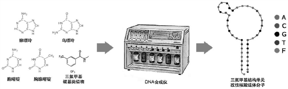
[0067] (1) if figure 1 As shown in the schematic diagram of the preparation method of the nucleic acid aptamer modified by the trifluoromethyl structural unit, the trifluoromethyl group is modified at both ends of the nucleic acid aptamer SGC8 by solid-phase synthesis, and simultaneously introduced into the 5' end of the nucleic acid aptamer The thiol group was used for further coupling, and the non-modified SGC8 nucleic acid aptamer was synthesized for control, and the specific sequence is as follows;
[0068] F-SGC8: 5’-(SH)-FFA TCT AAC TGC TGC GCC GCC GGG AAA ATA CTG TAC GGTTAG AFF-3’
[0069] SGC8: 5'-(SH)-ATC TAA CTG CTG CGC CGC CGG GAA AAT ACT GTA CGG TTA GA-3';
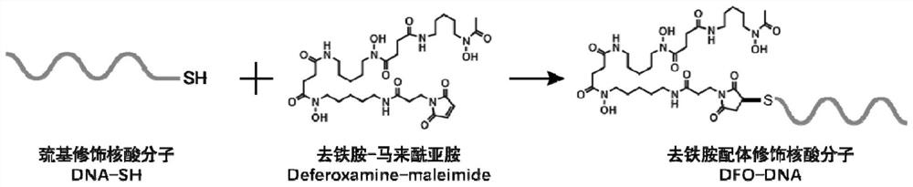
[0070] (2) if figure 2 As shown in the schematic diagram of the coupling of nucleic acid molecules and DFO ligands, 3 mg of DNA-SH synthesized in ste...
Embodiment 2
[0074]A tumor-bearing mouse model of PTK7 positive colorectal cancer was constructed. Specifically include the following steps:
[0075] (1) Use the PTK7-positive colorectal cancer tumor cell HCT116 cell line determined by Western blot to subculture in advance;
[0076] (2) The cultured HCT116 tumor cells were digested and resuspended in the cell culture medium, then mixed with Matrigel (Corning) at a ratio of 1:1, and the mixed cell suspension was placed on ice, Carry out tumor transplantation as soon as possible;
[0077] (3) Take 100 μl of the cell suspension in step 2 (which contains 2×10 6 cells), injected into the right axilla of 4–5 week old Balb / c nude mice (Victoria Lihua) to establish a subcutaneous xenograft tumor model.
Embodiment 3
[0079] Trifluoromethyl structure unit modified SGC8 nucleic acid aptamer ([ 89 Zr]DFO-SGC8-F) PET / CT molecular imaging of subcutaneous colorectal cancer. Specifically include the following steps:
[0080] (1) Inject 3.7–7.4 MBq into each tumor-bearing nude mouse via the tail vein[ 89 Zr]DFO-SGC8-F molecular probe (3–6 per group);
[0081] (2) At specific time points (3 days, 5 days and 7 days) after injection, use isoflurane (concentration: 3%) mixed with oxygen to anesthetize tumor-bearing nude mice, and put the nude mice into a state of deep anesthesia Place on the IRIS small animal PET / CT scanning bed in a prone position, continue to acquire PET and CT images, and use the software that comes with the IRIS system to complete image reconstruction. Such as Figure 7 as shown, 89 Zr labeled SGC8 aptamer ([ 89 Zr]DFO-SGC8) and trifluoromethyl structural unit modified SGC8 nucleic acid aptamer ([ 89 Zr]DFO-SGC8-F) PET / CT molecular images of tumor-bearing mice 3 days after ...
PUM
 Abstract
Description
Claims
Application Information
 - R&D
- Intellectual Property
- Life Sciences
- Materials
- Tech Scout
- Unparalleled Data Quality
- Higher Quality Content
- 60% Fewer Hallucinations
Browse by: Latest US Patents, China's latest patents, Technical Efficacy Thesaurus, Application Domain, Technology Topic, Popular Technical Reports.
© 2025 PatSnap. All rights reserved.Legal|Privacy policy|Modern Slavery Act Transparency Statement|Sitemap|About US| Contact US: help@patsnap.com