Method for differential diagnosis of EBV infected cell subtype and application thereof
A technology for infecting cells and differential diagnosis, applied in the medical field, can solve the problems of inconvenient tissue biopsy, high instrument requirements, poor sensitivity, etc., to ensure high sensitivity, high specificity, high clinical application value, reliable detection and identification. Effect
- Summary
- Abstract
- Description
- Claims
- Application Information
AI Technical Summary
Problems solved by technology
Method used
Image
Examples
Embodiment 1
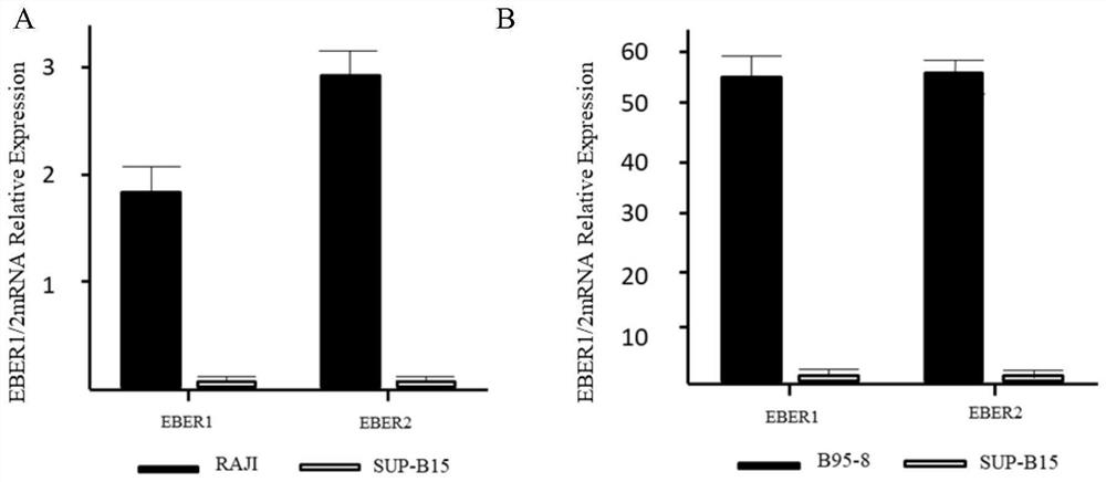
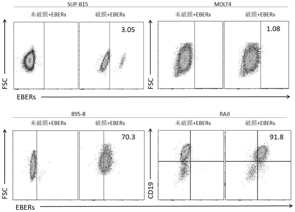
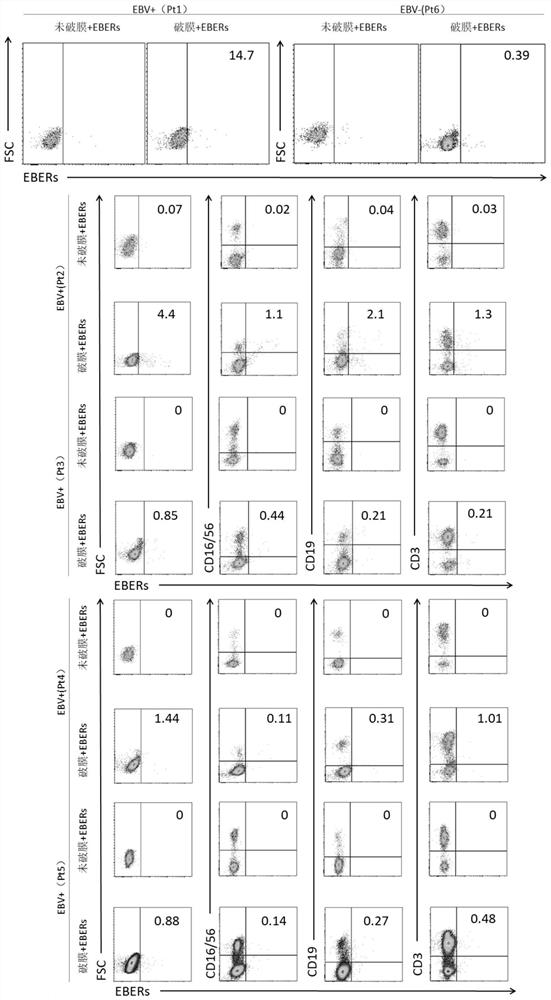
[0057] Application of Flow-FISH technology in cell lines and clinical patients
[0058] In this example, flow cytometry and fluorescence in situ hybridization are combined, and EBERs is used as the target RNA to hybridize with EBERs probes, and the expression of EBERs and surface antigens is detected simultaneously to identify the cell subtype infected with the virus. The specific experimental process is as follows:
[0059] 1. Research object
[0060] The research objects of this embodiment are EBV-negative and EBV-positive human cell lines cultured in vitro, and peripheral blood samples of clinical patients.
[0061] (1) EBV negative (EBV-) cell lines: human B lymphocytic leukemia cell line Sup-B15, human acute T cell leukemia cell line Molt-4.
[0062] EBV positive (EBV+) cell lines: marmoset EB virus transformed leukocyte line B95-8, human Burkitt's tumor cell line Raji.
[0063] The characteristics and sources of the above cell lines are shown in Table 2.
[0064] Tab...
Embodiment 2
[0108] Under normal circumstances, fluorescently labeled probes cannot enter the cell through the intact cell membrane. To measure RNA in the nucleus by flow cytometry, it is necessary to use a membrane-breaking agent to destroy the integrity of the cell membrane to create small holes for the probe to pass through. The effect of membrane disruption will directly affect the fluorescence staining and result analysis.
[0109] Based on this, referring to the experimental method of Example 1, this example explores the type of immobilized membrane-breaking agent suitable for flow cytometry-fluorescence in situ hybridization. The experimental method and process are as follows:
[0110] 1. By different membrane breaking agents: 0.2% TritonX-100, 0.2% Saponin, 0.2% Tween-20, and Thermo Fisher commercially available finished membrane breaking kits (TritonX-100, Saponin are all purchased from sigma-aldrich Sigma Aldrich (Shanghai) Trading Co., Ltd.; Tween-20 was purchased from Beijing S...
Embodiment 3
[0124] At present, there is no kit specifically for flow cytometry-fluorescence in situ hybridization on the market, all of which contain hybridization solutions (including 30% formamide and 10% dextran sulfate) designed for traditional fluorescence in situ hybridization. The state requirements are not high. In order to increase the fluorescence intensity of the probe, a high-concentration hybridization solution is usually used, but this has a great impact on the cell state, and it cannot be detected by flow cytometry when it is applied in the method of the present invention. Therefore, in this embodiment, the concentration of the main components (formamide and dextran sulfate) in the hybridization solution containing the probe was explored to find out the concentration of the hybridization solution suitable for flow cytometry-fluorescence in situ hybridization.
[0125] The main components of the hybridization solution used in this example are formamide and dextran sulfate. A...
PUM
 Abstract
Description
Claims
Application Information
 - R&D
- Intellectual Property
- Life Sciences
- Materials
- Tech Scout
- Unparalleled Data Quality
- Higher Quality Content
- 60% Fewer Hallucinations
Browse by: Latest US Patents, China's latest patents, Technical Efficacy Thesaurus, Application Domain, Technology Topic, Popular Technical Reports.
© 2025 PatSnap. All rights reserved.Legal|Privacy policy|Modern Slavery Act Transparency Statement|Sitemap|About US| Contact US: help@patsnap.com


