
Personalized 3D printing meniscus regeneration stent and preparation method thereof

A 3D printing and meniscus technology, applied in the field of medical prosthesis, can solve the problems of easy dislocation of meniscus bracket, large difference in heterogeneity distribution, poor cartilage protection effect of meniscus tissue, etc., to achieve enhanced regeneration and repair functions, Strengthen regeneration and repair, avoid dislocation and uneven stress distribution

Active Publication Date: 2022-02-01
PEKING UNIV THIRD HOSPITAL
View PDF10 Cites 0 Cited by
  • Summary
  • Abstract
  • Description
  • Claims
  • Application Information

AI Technical Summary

Problems solved by technology

However, most of the brackets produced in the previous research did not restore the shape of the original meniscus 1:1, and there were still some differences in shape with the natural meniscus, so they could not match the tibial plateau at the bottom and the femoral condyle above it, resulting in easy meniscus brackets. Dislocation, or uneven distribution of stress, ultimately affecting meniscal regeneration
Moreover, different growth factors can promote cells to produce different phenotypes and secrete different components. However, in previous studies, seed cells and growth factors were mostly evenly distributed throughout the scaffold, which eventually led to the regeneration of meniscus tissue in collagen composition, cell The phenotype is relatively single, which is quite different from the heterogeneous distribution of the natural meniscus, and cannot meet the functions of the natural meniscus
Ultimately, the regenerated tissue-engineered meniscus still has a large difference in collagen composition or distribution from the natural meniscus, or the regenerated meniscal tissue has poor cartilage protection

Method used


Image

  • Personalized 3D printing meniscus regeneration stent and preparation method thereof
  • Personalized 3D printing meniscus regeneration stent and preparation method thereof
  • Personalized 3D printing meniscus regeneration stent and preparation method thereof

Examples


Embodiment 1

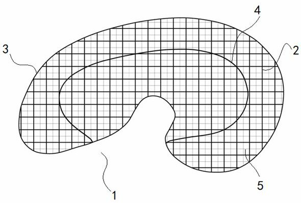
[0038] The purpose of this embodiment is to provide a method for preparing a personalized 3D printed meniscus regeneration bracket, including: preparing a bracket model and setting a support structure model for supporting the bracket model; The working parameters of the head and internal structure printing design; the extruded material in the high-temperature print head and the extruded material in the low-temperature print head are mixed and printed, and the inner 2 / 3 area of ​​​​the meniscus regeneration bracket is set as the "white area" and the outer 1 The / 3 area was set as the "red area" to construct the meniscus regeneration scaffold by partition printing.

[0039] The working principle of the above-mentioned technical scheme is:

[0040]Using bio-3D printing technology, the original meniscus-shaped scaffold is constructed at 1:1. At the same time, in order to make the prepared scaffold implantable, it can match the tibial plateau at the bottom and the femoral condyle a...

Embodiment 2

[0044] According to the preparation method of a personalized 3D printed meniscus regenerative scaffold described in Embodiment 1, in step S1, after obtaining the nuclear magnetic image data of the knee joint, the nuclear magnetic data of the knee joint is imported into the medical reverse software Mimics to construct a three-dimensional model of the meniscus, and then The constructed three-dimensional meniscus model was imported into Geomagic reverse engineering software for triangular surface optimization to obtain the scaffold model to be prepared. The detailed steps of this process are as follows:

[0045] 1. Knee joint MRI imaging and 3D model construction

[0046] Use a 3.0T nuclear magnetic resonance instrument to scan the knee joint to obtain the knee joint nuclear magnetic resonance image. The nuclear magnetic resonance scanning sequence is PD-FSE-SPIR, and the specific parameters are: repetition time (TR): 2915.0 milliseconds, echo time (TE): 39.4 millisecond. Then ...

Embodiment 3

[0055] According to the preparation method of a personalized 3D printed meniscus regeneration bracket described in Embodiment 2, in step S1, the support structure model is designed according to the bracket model, so that during 3D printing, there is an empty gap between the bracket model and the printing platform Filled by supporting structure model;

[0056] Wherein, the internal structure of the support structure model is the same as that of the model printed by the high temperature print head.

[0057] Specifically, Pluronic F127 is selected as the material for printing the support structure model. Pluronic F127 has the following typical advantages: 1. It can be printed at room temperature; 2. It has excellent rheological properties and high printing fidelity ;3. Pluronic F127 is a non-ionic surfactant that is soluble in water, so after printing, soak the meniscus bracket in water to completely remove the support structure;

[0058] The beneficial effect of the above techn...


PUM


Abstract

The invention provides a preparation method of a personalized 3D printing meniscus regeneration stent. The preparation method comprises the following steps: S1, a stent model is prepared, and a support structure model for support the stent model is set; s2, working parameters of a high-temperature printing head, a low-temperature printing head and internal structure printing design are set on a biological 3D printer; and S3, the extruded material in the high-temperature printing head and the extruded material in the low-temperature printing head are subjected to mixed printing, and the meniscus regeneration stent is constructed in a partition printing mode that the 2 / 3 area of the inner side of the meniscus regeneration stent is set as a white area, and the 1 / 3 area of the outer side of the meniscus regeneration stent is set as a red area. The personalized 3D printing meniscus regeneration stent has the advantages that the bottom of the personalized 3D printing meniscus regeneration stent can be matched with the tibial plateau; and the regeneration and repair functions after implantation can be effectively enhanced.

Description

technical field [0001] The invention relates to the technical field of medical prosthesis, in particular to a personalized 3D printed meniscus regeneration bracket and a preparation method thereof. Background technique [0002] The meniscus is a semilunar-shaped fibrocartilage tissue located between the femoral condyle and the tibial plateau cartilage. In the knee joint, it performs functions such as cushioning and decompression, absorbing shock, lubricating the joint, and maintaining joint stability. The blood supply of the joint capsule and synovium is called the "red zone". The inner 2 / 3 area lacks blood supply and only provides nutrition through the penetration of joint fluid, which is called the "white zone". [0003] As the number of people currently participating in sports continues to increase, the incidence of meniscus injuries is also increasing accordingly. Meniscal injury can significantly affect its function. Mild cases manifest as asymptomatic or mild joint pa...

Claims


Application Information

Patent Timeline
Patent Type & Authority Applications(China)
IPC IPC(8): A61F2/30C12M1/26B29C64/112B29C64/209B29C64/386B33Y50/00B33Y70/10B33Y80/00A61L27/18A61L27/22A61L27/38A61L27/54A61L27/56
CPCA61F2/30756A61F2/30942C12M33/00B29C64/112B29C64/386B29C64/209B33Y50/00B33Y70/10B33Y80/00A61L27/18A61L27/222A61L27/227A61L27/3834A61L27/3852A61L27/56A61L27/54A61F2002/30761A61F2002/30766A61F2002/30985A61F2002/30943A61L2430/06A61L2300/414A61L2300/30A61L2300/412
Inventor 闫文强敖英芳麦合木提·麦麦提敏程锦胡晓青
Owner PEKING UNIV THIRD HOSPITAL
Learn More