Preparation of a DNA quadrangular pyramid for visual detection of tumor-associated mRNAs in living cells
A tumor-related, quadrangular pyramid technology, applied in the field of biosensing, can solve the problems of time-consuming and labor-intensive, difficult to achieve real-time detection of single cells, and a large number of sample cells, so as to avoid false positive signals, save costs, and achieve good biocompatibility. Effect
- Summary
- Abstract
- Description
- Claims
- Application Information
AI Technical Summary
Problems solved by technology
Method used
Image
Examples
Embodiment 1
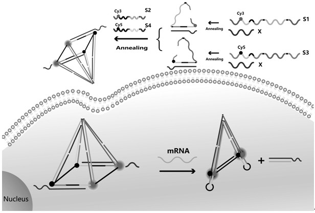
[0026] A method for preparing a DNA pyramid for visual detection of tumor-associated mRNA in living cells, comprising the following steps:
[0027] 1) Take equimolar S1 and CP, heat in a metal bath at 95ºC for 5 min, and naturally cool to room temperature to obtain mixture 1; then take equimolar S3 and CP to prepare as above to obtain mixture 2;
[0028] 2) After mixing mixture 1 and mixture 2, add equimolar S2 and S4, and anneal in the PCR instrument according to the following procedure: heat at 50ºC for 10 minutes, cool to 25ºC (3min / ºC), and hold at 4ºC for 60 minutes to obtain DNA pyramid at a final concentration of 1 μM;
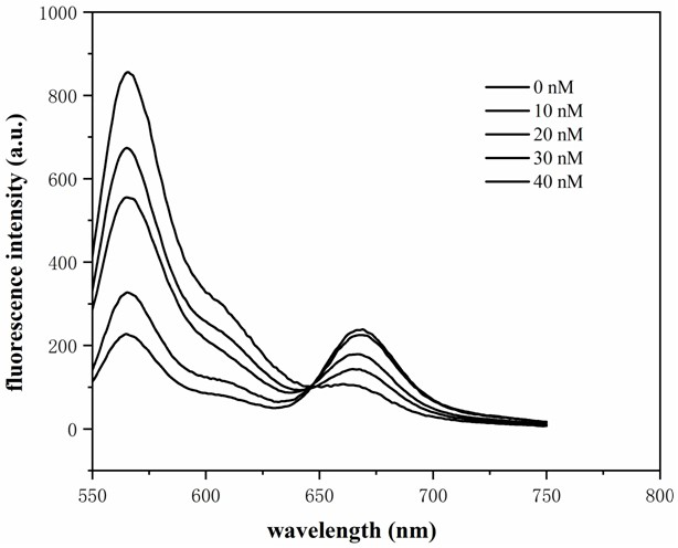
[0029] 3) Add different concentrations of c-myc target to measure fluorescence;
[0030] The specific operation method in step 3) is: in the buffer system containing 1×TAE / Mg2+, mix different amounts of the target substance with DNA at a concentration of 200 nM, so that the final concentrations of the target are 0, 10, 20, 30, 40 and 50 nM. Put all t...
Embodiment 2
[0032] A method for preparing a DNA pyramid for visual detection of tumor-associated mRNA in living cells, comprising the following steps:
[0033] Step 1), 2) are the same as in Example 1
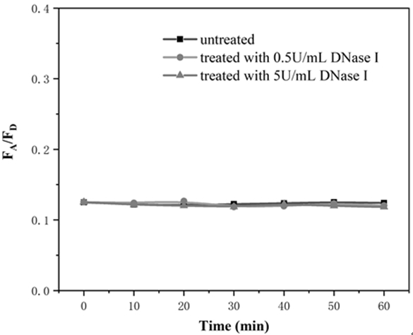
[0034] 3) The ability of DNA pyramids to avoid false positive signals. Add DNase I to the buffer containing DNA pyramids so that the final concentrations are 0.5 U / mL and 5 U / mL respectively, and incubate at 37 ºC in the dark for 0, After 10, 20, 30, 40, 50, and 60 min, measure the fluorescence intensity of the sample in the range of 540-570 nm under the excitation light wavelength of 525 nm, and the results are as follows image 3 shown. It is a well-established fact that DNA is easily degraded by nucleases ubiquitous in biological systems to generate false positive signals. Compared with single-intensity-based sensing probes, FRET-based fluorescence ratiometric signals can effectively avoid false positive signals due to nuclease digestion, protein binding, or thermodynamic fluctuations...
Embodiment 3
[0036] A method for preparing a DNA pyramid for visual detection of tumor-associated mRNA in living cells, comprising the following steps:
[0037] Step 1), 2) are the same as in Example 1
[0038] 3) In cell imaging experiments, MCF-7 cells were inoculated with DMEM medium in culture dishes and incubated in 5 % CO 2 Incubate for 24 hours at 37 ºC in an incubator. Add 100 nM of DNA to MCF-7 cells at 37 ºC and 5 % CO 2 Incubate for 4 h. Wash multiple times with PBS to remove excess sample. Under a confocal microscope, confocal fluorescence images of cells were obtained by laser scanning, the results are as follows Figure 4 shown. A clear FRET signal was observed in MCF-7 cells, indicating the presence of the target C-myc mRNA.
PUM
 Abstract
Description
Claims
Application Information
 - Generate Ideas
- Intellectual Property
- Life Sciences
- Materials
- Tech Scout
- Unparalleled Data Quality
- Higher Quality Content
- 60% Fewer Hallucinations
Browse by: Latest US Patents, China's latest patents, Technical Efficacy Thesaurus, Application Domain, Technology Topic, Popular Technical Reports.
© 2025 PatSnap. All rights reserved.Legal|Privacy policy|Modern Slavery Act Transparency Statement|Sitemap|About US| Contact US: help@patsnap.com


