
Fixing permeabilization wash buffer and preparation method thereof

A technology of fixative and membrane breaking agent, which is applied in the field of fixing membrane breaking agent and its preparation, which can solve the problems of large changes in cell light scattering, increase of experimental operation steps, and influence of detection results, and achieve simple operation, low detection cost, highly reproducible effect

Pending Publication Date: 2021-01-15
杭州联科生物技术股份有限公司
View PDF0 Cites 0 Cited by
  • Summary
  • Abstract
  • Description
  • Claims
  • Application Information

AI Technical Summary

Problems solved by technology

[0004] In the process of detecting intracellular cytokines by flow cytometry, whether the fixation and membrane rupture of the cells is appropriate has a great influence on the experimental results. At present, the membrane rupture agents used in many domestic hospitals and laboratories are mainly from foreign companies, and there is no such agent in China. Products comparable to foreign ones, but generally expensive. Domestic manufacturers fix the membrane breaking agent, which causes a large change in the light scattering of the cells after fixing the membrane breaking, which affects the detection results of the antigen expression level. The experimental conditions must be checked before the experiment. Optimization, adding experimental operation steps

Method used


Image

  • Fixing permeabilization wash buffer and preparation method thereof
  • Fixing permeabilization wash buffer and preparation method thereof
  • Fixing permeabilization wash buffer and preparation method thereof

Examples


Embodiment 1

[0035] A fixative, its composition and proportioning are:

[0036]

[0037] The pH of the fixative was 7.2.

[0038] A preparation method of a fixative, comprising the following steps:

[0039] Step 1: Accurately weigh sodium dihydrogen phosphate, dihydrate, disodium hydrogen phosphate, and sodium chloride into a beaker, add an appropriate amount of ultrapure water, stir and mix, and fully dissolve;

[0040] Step 2: Accurately weigh paraformaldehyde into a beaker, add it to the solution prepared in Step 1, heat and stir with a magnetic stirrer, keep the temperature below 60°C until it is completely dissolved, and adjust the pH value to 7.2 with 10N NaOH solution;

[0041] Step 3: After adjusting the pH, add ultrapure water into the beaker with a measuring cylinder, set the volume to the prepared volume, stir and mix well, and store at room temperature in the dark.

[0042] A membrane breaking agent, its composition and proportioning are:

[0043]

[0044]

[0045] ...

Embodiment 2

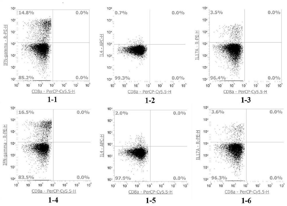
[0052] Such as figure 1 As shown, human Th1 / 2 / 17 flow detection:

[0053] Step 1: Add 250 μl of anticoagulated blood to the flow tube, add 250 μl of serum-free 1640 medium, vortex and mix, add 2 μl of PMA / Ionomycin Mixture (250×) and 2 μl of BFA / Monensin Mixture (250× ), vortex and mix well, take 250 μl of anticoagulant blood and add it to the flow tube, add 250 μl of serum-free 1640 medium as a negative control, vortex and mix well, and incubate for 4-6 hours in a carbon dioxide cell incubator , vortex and mix once every 1 hour;

[0054] Step 2: Take 100 μl cell suspension from the sample tube and control tube to a new flow tube, add 5 μl Anti-Human CD3, FITC and 5 μl Anti-HumanCD8α, PerCP-Cy5.5, vortex to mix, room temperature Incubate in the dark for 15 minutes;

[0055] Step 3: Add 100 μl of fixative to each tube, vortex to mix, and incubate at room temperature for 15 minutes in the dark;

[0056] Step 4: After the incubation, add 2ml 1×PBS to each tube, vortex to mix,...

Embodiment 3

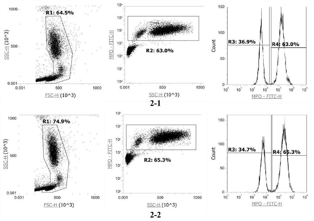
[0061]Step 1: Take 100 μl of anticoagulated blood and add it to the flow tube, add 100 μl of fixative, vortex to mix, and incubate at room temperature in the dark for 15 minutes;

[0062] Step 2: After the incubation, add 2ml 1×PBS to each tube, vortex to mix, centrifuge at 1500rpm for 5 minutes, and discard the supernatant;

[0063] Step 3: Add 100 μl membrane disrupting agent and 5 μl Anti-Human Myeloperoxidase (MPO), FITC to each tube, vortex to mix, and incubate at room temperature for 15 minutes in the dark;

[0064] Step 4: After the incubation, add 2ml 1×PBS to each tube, vortex to mix, centrifuge at 1500rpm for 5 minutes, and discard the supernatant;

[0065] Step 5: Add 500 μl 1×PBS to each tube to resuspend, and perform flow cytometry detection.


PUM


Abstract

The invention discloses a fixing agent. The fixing agent comprises 0.01-0.02 M of sodium dihydrogen phosphate dihydrate, 0.01-0.02 M of sodium dihydrogen phosphate, 0.05-0.15 M of sodium chloride, 4-6% of paraformaldehyde, and the balance of water. Further, the pH value of the fixing agent is 7.0-7.4. A preparation method of the fixing agent comprises the following steps: 1, accurately weighing sodium dihydrogen phosphate dihydrate, disodium hydrogen phosphate and sodium chloride into a beaker, adding a proper amount of ultrapure water, and uniformly stirring and mixing for sufficiently dissolving; 2, accurately weighing paraformaldehyde into a beaker, adding paraformaldehyde into the solution prepared in step 1, heating and stirring with a magnetic stirrer, controlling the temperature tobe 60 DEG C or below until paraformaldehyde is completely dissolved, and adjusting the pH value to 7.0-7.4 with a 10N NaOH solution; and 3, after the pH is adjusted, adding ultrapure water into the beaker by using a measuring cylinder, fixing the volume to a preparation volume, uniformly stirring and mixing, and storing in a dark place at room temperature. The advantages of good detection specificity and sensitivity, low detection cost, high stability, simple operation, high repeatability, and guaranteeing of the reliability of the result are achieved.

Description

technical field [0001] The invention relates to the technical field of flow cytometer detection, in particular to a fixed membrane breaking agent and a preparation method thereof. Background technique [0002] With the rapid development of flow cytometry, the application of flow cytometry combined with intracellular staining technology to detect intracellular cytokines is a new method established in recent years, which can simultaneously detect two or more cytokines at the single cell level , has been widely used due to its strong specificity, relatively fast and accurate quantification. [0003] When the immune response of immune cells is relatively weak, the level of secreted cytokines is difficult to be detected by the instrument, so stimulators are used to stimulate and activate lymphocytes in vitro, and then protein transport inhibitors are added to prevent cytokines from being secreted outside the cells. Stimulate activated cytokines to stay in the cell, and then fix ...

Claims


Application Information

Patent Timeline
Patent Type & Authority Applications(China)
IPC IPC(8): G01N1/30G01N15/14G01N33/68
CPCG01N1/30G01N15/14G01N33/6869G01N33/6866G01N2001/305G01N2333/5406G01N2333/54G01N2333/57
Inventor 郭爱龙
Owner 杭州联科生物技术股份有限公司
Learn More