Severe tumor image recognition system and method
A tumor imaging and recognition system technology, applied in neural learning methods, character and pattern recognition, biological neural network models, etc., can solve the problems of automatic tumor classification, grading and localization systems, and normal image discarding, and achieve automatic processing. The effect of high level, reduced workload and improved diagnosis speed
- Summary
- Abstract
- Description
- Claims
- Application Information
AI Technical Summary
Problems solved by technology
Method used
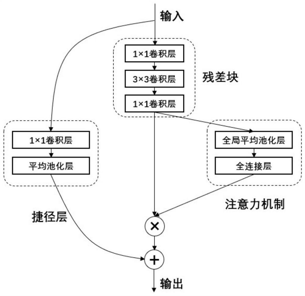
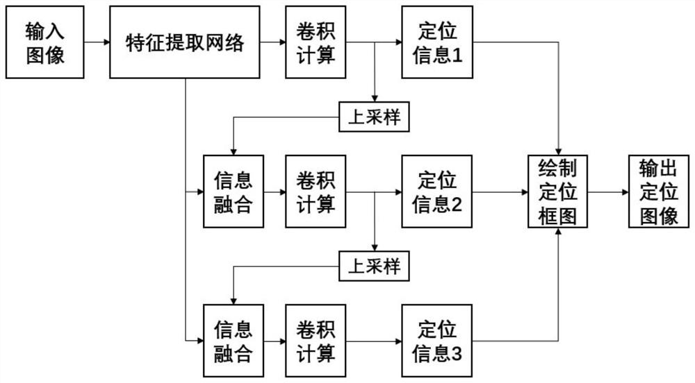
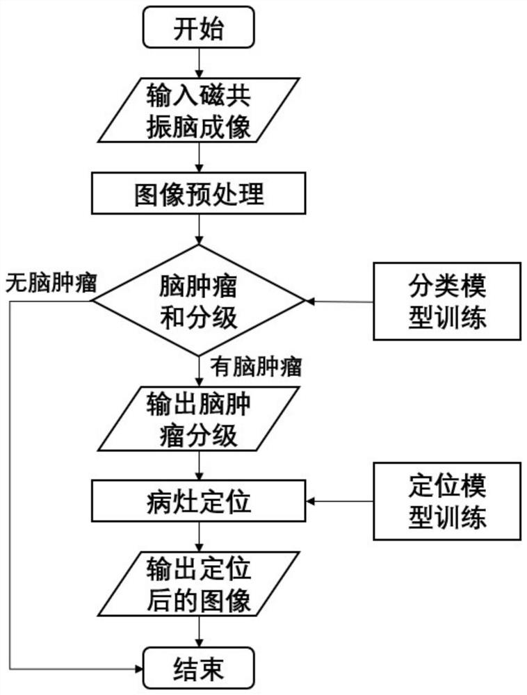
Image

Examples
Embodiment Construction
[0026] The present invention will be further elaborated below in conjunction with embodiment.
[0027]Step 1: Dataset collection and labeling. The present invention shares two data sets: classification data set and positioning data set. The present invention collects 957 magnetic resonance brain imaging images of different categories and levels from historical diagnostic case data, and divides them into 1) meningeal tumors (184 pictures) and 2) brain gliomas according to the manual diagnosis results of doctors Grade 1 (112 photos), 3) Glial Tumor Grade 2 (130 photos), 4) Glial Tumor Grade 3 (157 photos), 5) Glial Tumor Grade 4 (149 photos), 6) Anencephaly Tumors (225 photos), a total of 6 categories, were placed in the corresponding 6 folders, as the classification data set used in the first stage.
[0028] The image annotation tool Labelimg is used to annotate the tumor area in the images with tumors. The area where the tumor is located in each image is marked with a rectan...
PUM
 Abstract
Description
Claims
Application Information
 - R&D
- Intellectual Property
- Life Sciences
- Materials
- Tech Scout
- Unparalleled Data Quality
- Higher Quality Content
- 60% Fewer Hallucinations
Browse by: Latest US Patents, China's latest patents, Technical Efficacy Thesaurus, Application Domain, Technology Topic, Popular Technical Reports.
© 2025 PatSnap. All rights reserved.Legal|Privacy policy|Modern Slavery Act Transparency Statement|Sitemap|About US| Contact US: help@patsnap.com