SP-FCN-based MRI brain tumor image segmentation system and method
An image segmentation and brain tumor technology, applied in the field of MRI brain tumor image segmentation system based on SP-FCN, can solve the problems of artifact interference, low segmentation accuracy, long training time, etc. easy-to-capture effects
- Summary
- Abstract
- Description
- Claims
- Application Information
AI Technical Summary
Problems solved by technology
Method used
Image

Examples
Embodiment 1
[0036] The purpose of this embodiment is to provide an MRI brain tumor segmentation method based on SP-FCN.
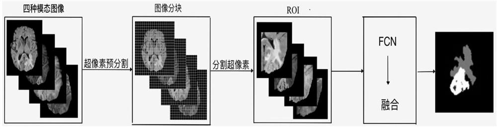
[0037] The data set selected in this implementation case has a total of 400 samples, the total number of samples used for training is 300, and the total number of samples used for testing is 100, such as figure 1 As shown, the overall process of the MRI brain tumor segmentation algorithm based on SP-FCN is shown, an MRI brain tumor segmentation method based on SP-FCN, including:
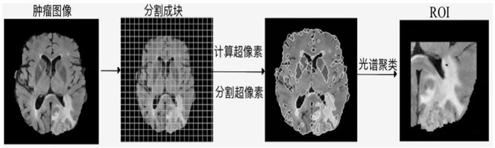
[0038] Obtain MRI brain tumor images of different modalities of the same sample, and divide the brain tumor image area into image blocks of equal size;
[0039] performing superpixel calculation on the image block;
[0040] performing spectral clustering based on the superpixel results of the image block, and segmenting the superpixels of the image block to obtain tumor superpixels and non-tumor superpixels;
[0041] Identify the image block where the tumor superpixel is located in the orig...
Embodiment 2
[0100] The purpose of this embodiment is to provide an MRI brain tumor segmentation system based on SP-FCN.
[0101] An MRI brain tumor segmentation system based on SP-FCN, including:
[0102] The image acquisition module is used to acquire MRI brain tumor images of the same sample with different modalities by using a nuclear magnetic resonance instrument, and divide the brain tumor image area into image blocks of equal size;
[0103] A superpixel calculation module, configured to perform superpixel calculation on the image block;
[0104] The superpixel segmentation module is used to perform spectral clustering based on the superpixel results of the image block, segment the superpixels, and obtain tumor superpixels and non-tumor superpixels;
[0105] The tumor image recognition module is used to identify the image block where the tumor superpixel is located in the original MRI brain tumor image, and obtain ROI images of MRI brain tumor images with different modalities;
[0...
Embodiment 3
[0162] The purpose of this embodiment is to provide an electronic device.
[0163]An electronic device, comprising, a memory, a processor, and a computer program stored on the memory, and the processor implements the following steps when executing the program, including:
[0164] Obtain MRI brain tumor images of different modalities of the same sample, and divide the brain tumor image area into image blocks of equal size;
[0165] performing superpixel calculation on the image block;
[0166] performing spectral clustering based on the superpixel results of the image block, and segmenting the superpixels of the image block to obtain tumor superpixels and non-tumor superpixels;
[0167] Identify the image block where the tumor superpixel is located in the original MRI brain tumor image, and obtain the ROI of the MRI brain tumor image with different modalities;
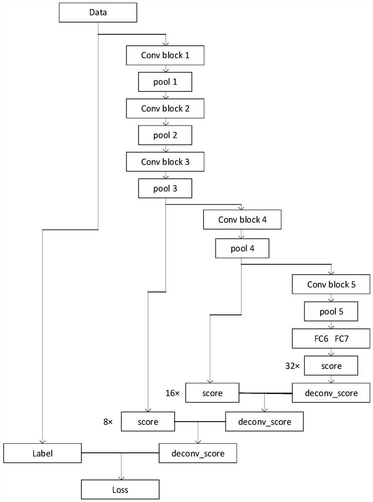
[0168] The ROIs of different modalities were input into the FCN network model for image fusion to obtain the tumor ...
PUM
 Abstract
Description
Claims
Application Information
 - R&D
- Intellectual Property
- Life Sciences
- Materials
- Tech Scout
- Unparalleled Data Quality
- Higher Quality Content
- 60% Fewer Hallucinations
Browse by: Latest US Patents, China's latest patents, Technical Efficacy Thesaurus, Application Domain, Technology Topic, Popular Technical Reports.
© 2025 PatSnap. All rights reserved.Legal|Privacy policy|Modern Slavery Act Transparency Statement|Sitemap|About US| Contact US: help@patsnap.com