Polypeptide Raman probe for targeted recognition of collagen and preparation method of polypeptide Raman probe
A collagen and target recognition technology, applied in Raman scattering, measuring devices, instruments, etc., can solve the problems of inaccurate positioning, non-specific adsorption, etc., and achieve narrow Raman slit width, good stability, and anti-photobleaching ability strong effect
- Summary
- Abstract
- Description
- Claims
- Application Information
AI Technical Summary
Problems solved by technology
Method used
Image
Examples
Embodiment 1
[0044] Example 1 Polypeptide Raman probe Cys-Ahx-(Gly-Pro-Hyp) 7 - Preparation of Ag nanoparticles-4-MBN
[0045] The designed peptide Raman probe is: Cys-Ahx-(Gly-Pro-Hyp) 7 -Ag nanoparticles-4-MBN.
[0046] Preparation of peptide Raman probes:
[0047] (1) Solid phase synthesis of collagen targeting polypeptide Cys-Ahx-(Gly-Pro-Hyp) 7 :
[0048] ① Add 100mg of Rink ammonia resin to a reactor with a sieve plate, and use 5mL of dichloromethane to swell the resin;
[0049] ②The N-terminal Fmoc protecting group was removed by 20% piperidine / N,N-dimethylformamide (DMF) solution, and the protecting group was completely removed by color reaction detection;
[0050] ③ Dissolve the amino acid (4eq) protected by Fmoc at the N-terminal, HOBt (4eq) and HBTU (4eq) in DMF, activate at low temperature for 20 minutes, add DIEA (6eq) dropwise to the solution, mix the solution and add it to the reactor, Response 3hrs.
[0051] ④ After the reaction, the reaction solution was extracted f...
Embodiment 2
[0057] Example 2 Preparation of Polypeptide Raman Probe Cys-Ahx-LRELHLNNNG-Ag Nanoparticle-R1
[0058] The designed polypeptide Raman probe is: Cys-Ahx-LRELHLNNNG-Ag nanoparticle-R1, and the R1 is S-(4-trimethylsilylethynyl-phenyl) thioacetate.
[0059] Preparation of peptide Raman probes:
[0060] (1) Solid phase synthesis of collagen targeting polypeptide Cys-Ahx-LRELHLNNNG:
[0061] ① Add 100mg of Rink ammonia resin to a reactor with a sieve plate, and use 5mL of dichloromethane to swell the resin;
[0062] ②The N-terminal Fmoc protecting group was removed by 20% piperidine / N,N-dimethylformamide (DMF) solution, and the protecting group was completely removed by color reaction detection;
[0063] ③ Dissolve the amino acid (4eq) protected by Fmoc at the N-terminal, HOBt (4eq) and HBTU (4eq) in DMF, activate at low temperature for 20 minutes, add DIEA (6eq) dropwise to the solution, mix the solution and add it to the reactor, Response 3hrs.
[0064] ④ After the reaction, t...
Embodiment 4
[0083] Example 4 In vitro tissue imaging of two kinds of polypeptide Raman probes
[0084] 1. Raman probe Cys-Ahx-(Gly-Pro-Hyp) 7 -Ag nanoparticles-4-MBN tissue imaging in vitro
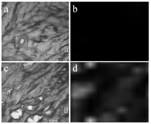
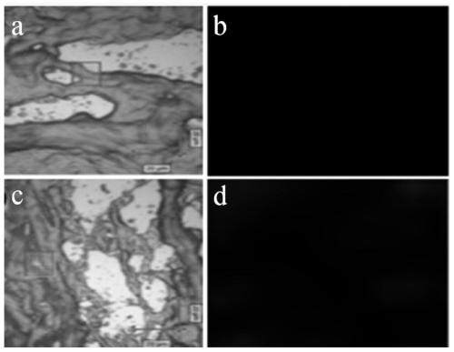
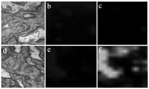
[0085] Add 100 μL, 400 pM polypeptide Raman probe Cys-Ahx-(Gly-Pro-Hyp) prepared in Example 1 to the frozen section of rat tail tissue 7 -Ag nanoparticles-4-MBN solution and the random probe Cys-Ahx-PGGOOPPGGOOPPGGOOPPGOG-Ag nanoparticles-4-MBN prepared in Comparative Example 1 were stained, and incubated at 4° C. for 2 hrs. After aspirating the staining solution, wash 5 times with 1×PBS to wash away unbound probes. Using a Raman microscope to observe and take pictures, the results are as follows figure 1 Shown, where a is the bright field image of the random probe Cys-Ahx-PGGOOPPGGOOPPGGOOPPGOG-Ag nanoparticles-4-MBN on rat tail tissue staining; b is the random probe Cys-Ahx-PGGOOPPGGOOPPGGOOPPGOG-Ag nanoparticles-4- Dark field image of rat tail tissue stained by MBN; c is polypeptide Raman prob...
PUM
 Abstract
Description
Claims
Application Information
 - R&D
- Intellectual Property
- Life Sciences
- Materials
- Tech Scout
- Unparalleled Data Quality
- Higher Quality Content
- 60% Fewer Hallucinations
Browse by: Latest US Patents, China's latest patents, Technical Efficacy Thesaurus, Application Domain, Technology Topic, Popular Technical Reports.
© 2025 PatSnap. All rights reserved.Legal|Privacy policy|Modern Slavery Act Transparency Statement|Sitemap|About US| Contact US: help@patsnap.com


