Preparation method of zin-ion-containing tissue engineering scaffold with anti-inflammatory function

A tissue engineering scaffold, zinc ion technology, applied in the field of biomedical engineering, can solve the problems of incomparable biomechanical and physiological functions of hyaline cartilage, unsatisfactory treatment effect, unable to meet functional requirements, etc., and achieve a good internal immune environment, Good histocompatibility, good cell adhesion

Active Publication Date: 2020-04-17
SHANDONG FIRST MEDICAL UNIV & SHANDONG ACADEMY OF MEDICAL SCI
View PDF4 Cites 3 Cited by
  • Summary
  • Abstract
  • Description
  • Claims
  • Application Information

AI Technical Summary

Problems solved by technology

Microfracture and subchondral bone drilling have been used successively for the treatment of cartilage injuries, but these treatment methods also face many problems, the most important of which is that the repaired cartilage is usually fibrocartilage, and its biomechanics and physiological functions are similar to those of hyaline cartilage. Incomparable, unable to meet functional requirements
Secondly, the additional damage to the non-weight-bearing area and the insufficient expansion of the cartilage damage area make the therapeutic effect of the above treatment methods unsatisfactory.

Method used


Image

  • Preparation method of zin-ion-containing tissue engineering scaffold with anti-inflammatory function
  • Preparation method of zin-ion-containing tissue engineering scaffold with anti-inflammatory function
  • Preparation method of zin-ion-containing tissue engineering scaffold with anti-inflammatory function

Examples


Embodiment 1

[0037] Embodiment 1 Preparation of simple Wharton's gum stent

[0038] (1) After taking out the umbilical cord, wash away the remaining blood in and on the surface of the umbilical vessels with normal saline, remove the outer capsule and internal blood vessels of the umbilical cord on a sterile operating table, and the separated transparent jelly-like substance is the human umbilical cord Wharton glue;

[0039](2) Repeatedly wash the Wharton gum twice with distilled water, then place the Wharton gum in the pulverizer, add triple distilled water, start the pulverization, and repeat several times until the Wharton gum is in a homogenized state;

[0040] (3) Put the Wharton gum homogenate prepared in step (2) into a refrigerator for freezing, then thaw at room temperature, and freeze and thaw repeatedly 3 to 4 times;

[0041] (4) Use gradient centrifugation to take the supernatant, add 1% TritonX-100 (polyethylene glycol octylphenyl ether), 0.25% trypsin, Tris-HCl buffer in sequ...

Embodiment 2

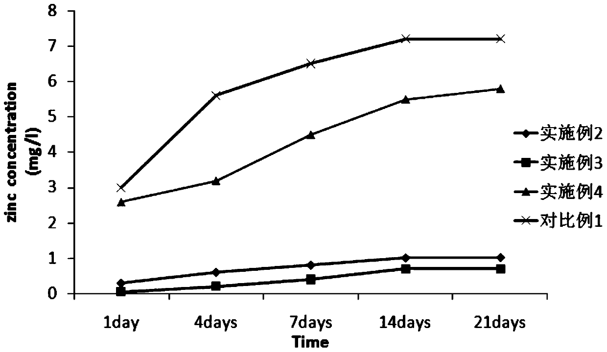
[0044] (1) Prepare a simple Wharton's gum stent according to the method described in steps (1) to (5) in Example 1;

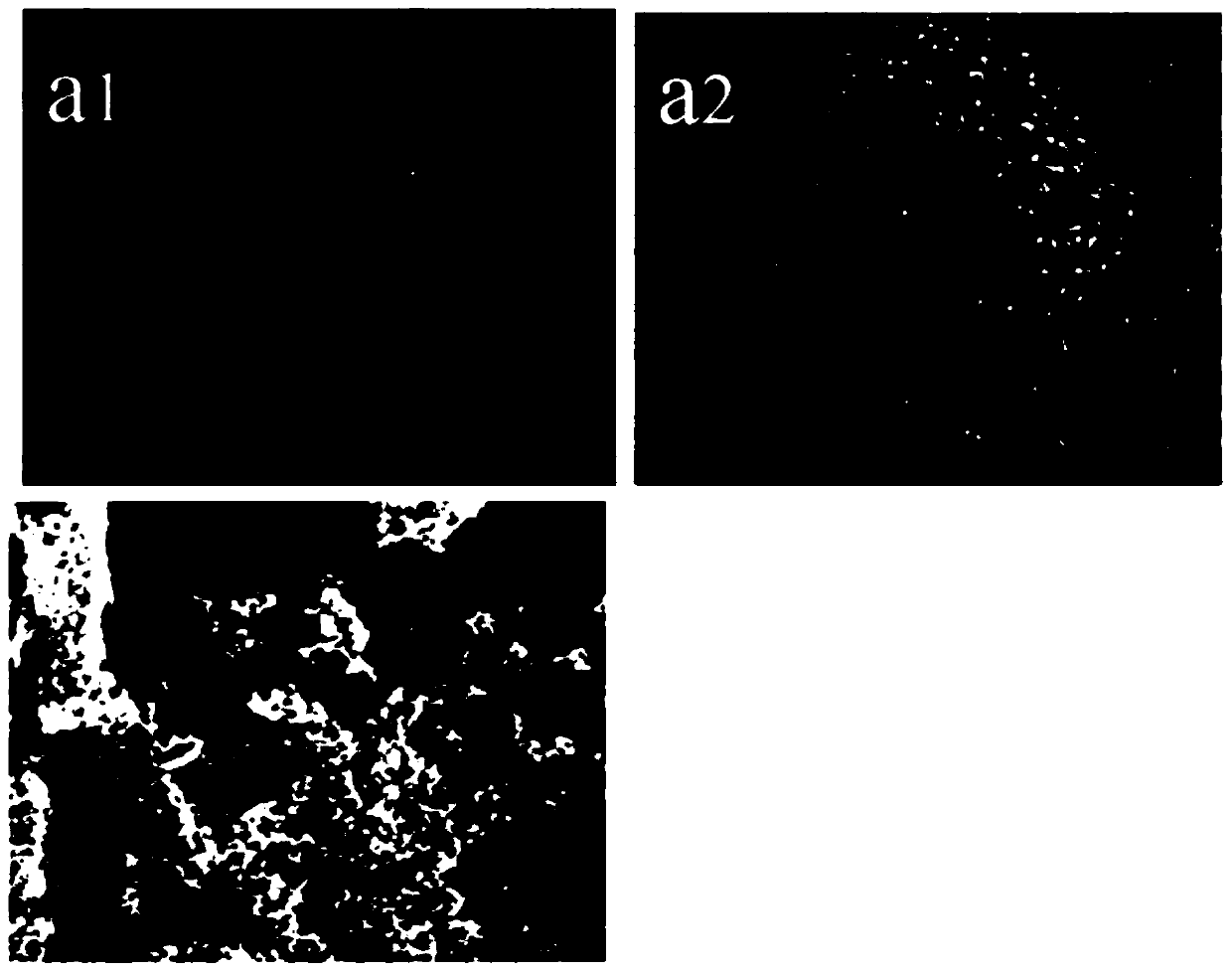
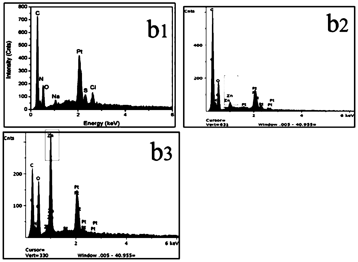
[0045] (2) Take NaCl: 120mmol / L, NaHCO respectively 3 : 2.0mmol / L, KCl: 4.0mmol / L, K 2 HPO 4 ·3H 2 O: 3.0mmol / L, HCl: 40.0mmol / L, CaCl 2 : 3.0mmol / L, Na 2 SO 4 : 0.8mmol / L, (CH 2 OH)3CNH 2 (Tris): 40mmol / L, Zn(CH 3 COO) 2 : 10mmol / L, prepare zinc-containing simulated body fluid, and place the simple Wharton gum stent prepared in step (1) in the simulated body fluid to induce deposition for 24 hours at 37°C;

[0046] (3) Take out the Wharton gum stent soaked in step (2), place it in a 5% ammonia solution in a hydrothermal kettle, and perform a gas phase hydrothermal reaction at 50°C for 24 hours;

[0047] (4) Place the Wharton gum support loaded with zinc ions prepared in step (3) in a vacuum freeze dryer, then place it in a 60°C irradiation sterilizer for irradiation disinfection, and place it at 4°C Store under airtight conditions in the refrigerato...

Embodiment 3

[0049] (1) Prepare a simple Wharton's gum stent according to the method described in steps (1) to (5) in Example 1;

[0050] (2) Take NaCl: 137.0mmol / L, NaHCO respectively 3 : 4.0mmol / L, KCl: 3.6mmol / L, K 2 HPO 4 ·3H 2 O: 4.0mmol / L, HCl: 30.0mmol / L, CaCl 2 : 2.5mmol / L, Na 2 SO 4 : 0.2mmol / L, (CH 2 OH)3CNH 2 (Tris): 30.0mmol / L, Zn(CH 3 COO) 2 : 5.0mmol / L zinc-containing simulated body fluid was prepared, and the simple Wharton's gum stent prepared in step (1) was placed in the simulated body fluid to induce deposition at 37°C for 24 hours;

[0051] (3) Take out the Wharton gum stent soaked in step (2), place it in a hydrothermal kettle with a concentration of 5% ammonia solution, and perform a gas phase hydrothermal reaction at 70°C for 48 hours;

[0052] (4) Place the Wharton gum support loaded with zinc ions prepared in step (3) in a vacuum freeze dryer, then place it in a 60°C irradiation sterilizer for irradiation disinfection, and place it at 4°C Store under air...


PUM


Abstract

The invention discloses a preparation method of a zin-ion-containing tissue engineering scaffold with an anti-inflammatory function. The method comprises the following steps: separating Wharton's jelly from a human umbilical cord; preparing a pure Wharton's jelly scaffold through the processes of repeated low-temperature crushing, freeze thawing, gradient centrifugation, pancreatin digestion and elution, vacuum freeze compression drying and the like; performing induced deposition on the pure Wharton jelly scaffold in simulated body fluid containing zinc ions; putting the scaffold in an ammoniawater solution in a hydrothermal kettle and performing a gas-phase hydrothermal reaction; and then performing vacuum freeze drying to obtain the cartilage tissue engineering scaffold containing zincions. The tissue engineering scaffold prepared by the method not only has high histocompatibility but also can successfully load zinc ions and release the zinc ions through a slow release effect, so that macrophage polarization is regulated and an anti-inflammatory effect on cartilage injury in an inflammatory environment is realized; and stem cells can be induced to differentiate into chondrocytes; and a good internal immune environment is provided for cartilage repair.

Description

【Technical field】 [0001] The invention relates to the technical field of biomedical engineering, in particular to a preparation method of a zinc ion-containing tissue engineering scaffold with anti-inflammatory function. 【Background technique】 [0002] Due to the lack of nutritional support of blood vessels and nerves, the regeneration ability and self-healing ability of articular cartilage are poor, so that it is very difficult to repair articular cartilage after injury. The current treatment of full-thickness articular cartilage injuries has been a great challenge for orthopedics and sports medicine doctors. Microfracture and subchondral bone drilling have been used successively for the treatment of cartilage injuries, but these treatment methods also face many problems, the most important of which is that the repaired cartilage is usually fibrocartilage, and its biomechanics and physiological functions are similar to those of hyaline cartilage. Incomparable, unable to me...

Claims


Application Information

Patent Timeline
IPC IPC(8): A61L27/36A61L27/04A61L27/50A61L27/54
CPCA61L27/3687A61L27/3691A61L27/3654A61L27/047A61L27/50A61L27/54A61L2300/41A61L2430/06A61L2300/102A61L2300/412A61L2430/40
Inventor 亓建洪毕懿康
Owner SHANDONG FIRST MEDICAL UNIV & SHANDONG ACADEMY OF MEDICAL SCI
Why Eureka
Try Eureka