
Chest X-ray pneumothorax segmentation method based on deep learning

A deep learning and chest technology, applied in the field of image processing, can solve the problems of delayed disease, blind spots in pneumothorax segmentation technology, and the inability of doctors to concentrate, and achieve the effect of improving segmentation accuracy.

Inactive Publication Date: 2020-03-20
SOUTHWEAT UNIV OF SCI & TECH
View PDF3 Cites 5 Cited by
  • Summary
  • Abstract
  • Description
  • Claims
  • Application Information

AI Technical Summary

Problems solved by technology

Due to the accumulation of a large number of chest X-rays, those patients with critical diseases such as pneumothorax may wait for a long time on the list, thus delaying the treatment
At the same time, long hours of overloaded work prevent doctors from concentrating
[0004] Most of the current research is based on deep learning pneumothorax recognition methods, but there are still blind spots in the pneumothorax segmentation technology for chest X-ray

Method used


Image

  • Chest X-ray pneumothorax segmentation method based on deep learning
  • Chest X-ray pneumothorax segmentation method based on deep learning
  • Chest X-ray pneumothorax segmentation method based on deep learning

Examples


Embodiment

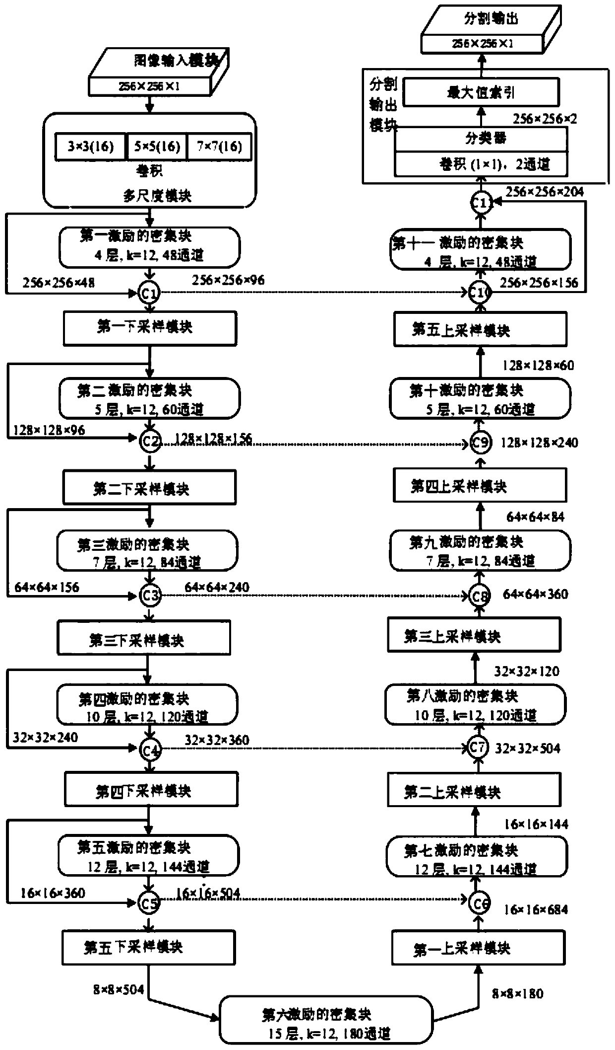
[0052] With the development of deep learning, the successful application of neural networks represented by convolutional neural networks in the field of computer vision has laid a foundation for the application of fully convolutional neural networks in medical image lesion segmentation. The fully convolutional neural network generally extracts features from the downsampling path, restores the image resolution through the upsampling path, and automatically learns the feature map from the original input to the expected output. Compared with the complex feature extraction process of traditional algorithms, the convolutional neural network is at an advanced The ability of abstract feature extraction is more significant, especially for the recognition of fine-grained images, which has great advantages and potential.

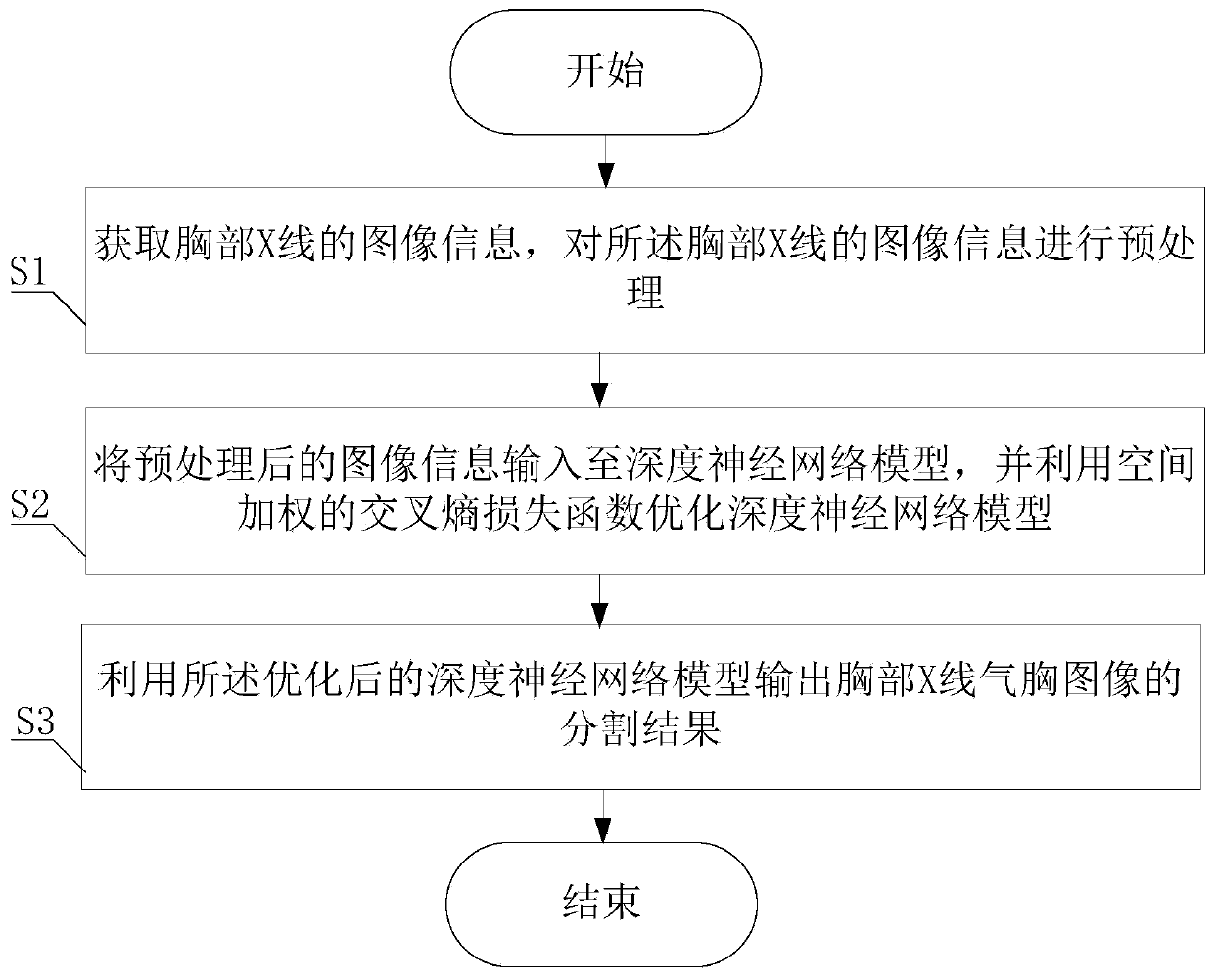
[0053] The embodiment of the present application provides a chest X-ray pneumothorax segmentation method based on deep learning, which is used to improve the accuracy ...


PUM


Abstract

The invention provides a chest X-ray pneumothorax segmentation method based on deep learning, and the method comprises the steps: obtaining the image information of chest X-rays, and carrying out thepreprocessing of the image information of the chest X-rays; inputting the preprocessed image information into a deep neural network model, and optimizing the deep neural network model by using a spatially weighted cross entropy loss function; and outputting a segmentation result of the chest X-ray pneumothorax image by utilizing the optimized deep neural network model. According to the method, anend-to-end deep neural network model is trained, and the deep neural network model finds out imaging features depicting pneumothorax through continuous autonomous learning so as to segment out suspected pneumothorax regions. Accurate segmentation of chest X-ray pneumothorax is realized, and accurate pneumothorax segmentation can provide important reference for subsequent treatment of a patient.

Description

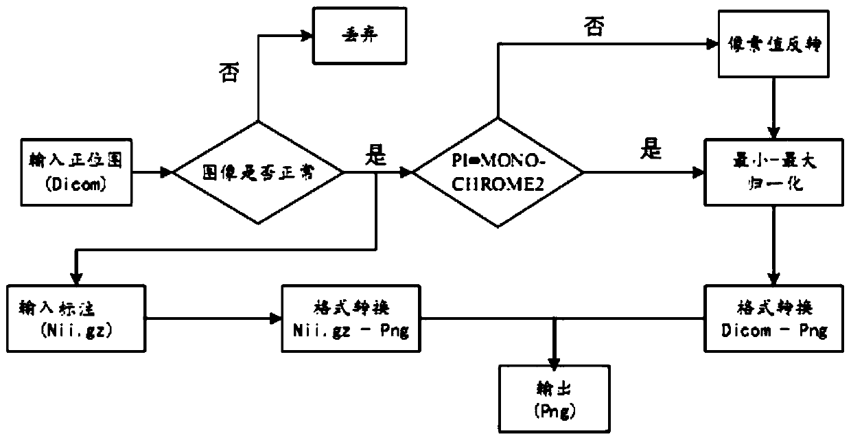
technical field [0001] The invention belongs to the technical field of image processing, and in particular relates to a chest X-ray pneumothorax segmentation method based on deep learning. Background technique [0002] Pneumothorax refers to air entering the pleural cavity, causing pneumothorax and lung collapse. It is a high-risk disease among lung diseases. The lung tissue and visceral pleura are usually ruptured due to lung disease or external force, or the tiny emphysema bubbles near the lung surface are ruptured, and the air in the lungs and bronchi escapes into the pleural cavity. If it is not treated in time, once it develops into hemopneumothorax or tension pneumothorax, it will pose a great threat to the patient's life, and the purpose of active treatment measures is to remove the gas in the chest cavity as soon as possible, so that the atrophied lung can be re-expanded as soon as possible, and the lung function can be restored. . [0003] Chest X-ray-based imagin...

Claims


Application Information

Patent Timeline
Patent Type & Authority Applications(China)
IPC IPC(8): G06T7/10G06N3/04G06N3/08
CPCG06T7/10G06N3/08G06T2207/10116G06N3/045
Inventor 罗国婷刘志勤王庆凤刘启榆周莹郑介志黄俊徐卫云
Owner SOUTHWEAT UNIV OF SCI & TECH
Learn More