Cell strain subjected to long-term passage after gamma ray irradiation of human skin fibroblasts and construction method of cell strain
A technology of fibroblasts and gamma rays, applied in the field of biomedicine, can solve the problems of not being able to better reveal the reasons for changes in the pathological mechanism of cell irradiation, the method of cell model construction has not been studied in depth, and the value of cells is limited. The effect of high scientific research and production application value
- Summary
- Abstract
- Description
- Claims
- Application Information
AI Technical Summary
Problems solved by technology
Method used
Image
Examples
Embodiment 1
[0025] Example 1: Establishment and verification of stable long-term passage human skin fibroblast (HFF-1) cells after irradiation
[0026] Human skin fibroblast (HFF-1) cells were recovered and cultured in DMEM medium containing 10% fetal bovine serum (FBS) and 1% penicillin-streptomycin dual antibiotic solution at 37°C. , 5% CO 2 Cultivate in an incubator; when the degree of cell mixing reaches 90%, use 1ml of trypsin containing EDTA (the mass concentration of EDTA is 0.25%) to digest, after digesting for 5min, use 2ml of culture medium to stop the digestion of cells in the culture flask, and put the culture flask After the 3ml liquid in the medium was repeatedly pipetted evenly, it was transferred into a 15ml centrifuge tube and placed at a Co60γ-ray source for irradiation, with a total irradiation dose of 10Gy. After the irradiated cells were taken back to the cell room, they were laid flat in the culture flask and continued to be cultured. After the cells reached more th...
Embodiment 2
[0027] Example 2: Morphological observation of long-term passage human skin fibroblast (HFF-1) cells after irradiation
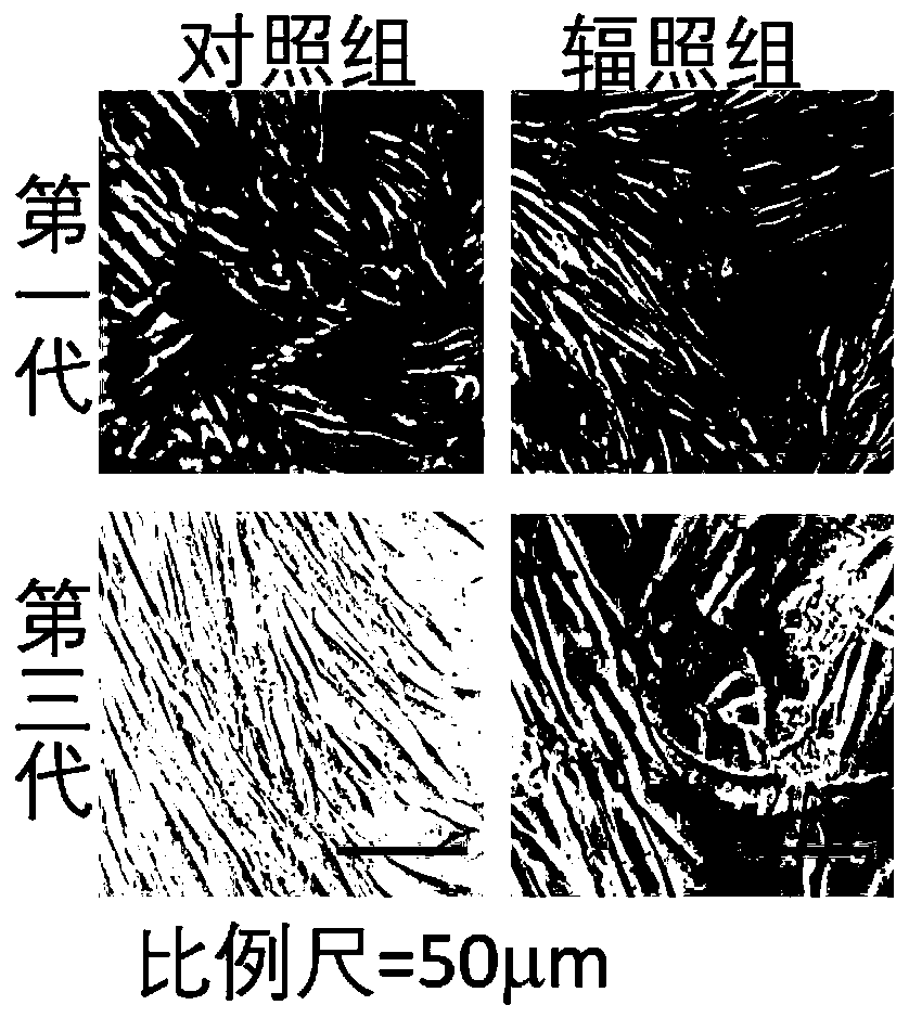
[0028] Inverted phase-contrast microscope was used to observe the morphology of living cells: human skin fibroblasts (HFF-1) control cells in the logarithmic growth phase and human skin fibroblasts (HFF-1) irradiated cells were taken respectively, washed with normal saline and replaced in the The living cell morphology of the first and third passage cells was observed under an inverted microscope (×200 times). The cell morphology of the first generation of cells did not change significantly after gamma ray irradiation. The density gradually decreased, the boundary was blurred, irregular vesicles appeared in the cytoplasm of some cells, and the cell membrane became transparent and bright, showing a pre-apoptotic shape ( figure 2 ).
Embodiment 3
[0029] Embodiment 3: Determination of the proliferation curve curve of long-term passage human skin fibroblast (HFF-1) cells
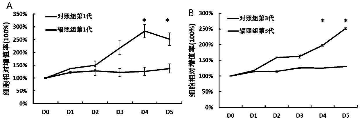
[0030] Normal control cells and irradiated mouse embryonic fibroblasts were digested with trypsin and then added a suitable new medium to resuspend the cells, according to 5×10 3 The concentration of cells / ml was inoculated into a 96-well plate, and each of the control group, the first generation and the third generation of the irradiation group had 5 controls to ensure that the cells were evenly dispersed in each culture well, and placed in the incubator overnight , to be detected after it adheres to the wall. The method is to add 10 μl of CCK-8 solution to each well, incubate in a 37°C incubator for 1 hour, detect the absorbance OD value at a wavelength of 476nm, and measure continuously for 5-7 days according to the actual growth of the cells. SPSS 19.0 software system was used for statistical analysis, and P﹤0.05 was considered statistically signi...
PUM
 Abstract
Description
Claims
Application Information
 - R&D Engineer
- R&D Manager
- IP Professional
- Industry Leading Data Capabilities
- Powerful AI technology
- Patent DNA Extraction
Browse by: Latest US Patents, China's latest patents, Technical Efficacy Thesaurus, Application Domain, Technology Topic, Popular Technical Reports.
© 2024 PatSnap. All rights reserved.Legal|Privacy policy|Modern Slavery Act Transparency Statement|Sitemap|About US| Contact US: help@patsnap.com


