A kind of human normal corneal epithelial cell and its application
A technology of corneal epithelial cells and epithelial cells, applied in the field of cell biology, to achieve the effect of tight arrangement, strong three-dimensional effect, and clear cell boundaries
- Summary
- Abstract
- Description
- Claims
- Application Information
AI Technical Summary
Problems solved by technology
Method used
Image
Examples
Embodiment 1
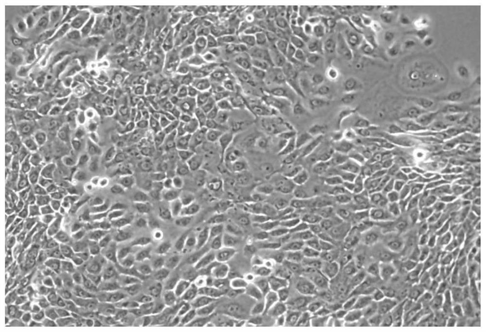
[0046] [Example 1] Primary isolation and culture of primary human normal corneal epithelial cells
[0047] (1) With the informed consent of the patient or patient guardian, collect the normal tissue samples next to the pterygium surgically removed from patients with pterygium.
[0048] (2) Preparation of digestive solution: HL medium containing 0.2 mg / mL of collagenase and dispase; among them, HL medium is: DMEM (GIBCO#11965-092) and serum-free medium SFM (GIBCO#10744- 019) mix by volume ratio 1:3, add the fetal calf serum of 5% (v / v) simultaneously, and 0.4 μ g / mL cortisol (hydrocortisone), 5 μ g / mL insulin (ins μ Lin), 8.4 ng / mL cholera toxin ( cholera toxin), 10ng / mL epithelial growth factor (EGF), 24μg / mL adenine, 100U / mL penicillin, 100μg / mL streptomycin, 0.25μg / mL amphoteric Mycin B (Fungizone), 30 μM Fasudil (Fasudil), the above medium needs to be filtered through a 0.22 μm pore size filter).
[0049] (3) Wash the isolated tissue sample once with 95-100% (v / v) ethanol...
Embodiment 2
[0058] [Example 2] Subculture of normal human corneal epithelial cells
[0059] (1) When the human normal corneal epithelial cells cultured in T25 or T75 culture flask proliferate to 70-90% abundance, wash the cells twice with 1×PBS (0.01M, pH 7.4), and then wash with 0.05% ( Mass volume ratio) Trypsin-EDTA digested monolayer cells for 2-5 minutes.
[0060] (2) Add 10 mL of complete DMEM to neutralize the digestion reaction for 1-2 minutes.
[0061] (3) Centrifuge at 1000rmp for 5 minutes, remove the supernatant, resuspend the cell pellet and inoculate in 10mL HL medium.
[0062] (4) If necessary, 1×10 6 The epithelial cells were resuspended in 1-2 mL of cell freezing solution (90% fetal bovine serum and 10% DMSO, v / v), and stored in liquid nitrogen for future use.
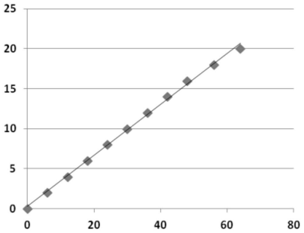
[0063] Subculture human normal corneal epithelial cells according to the above method, and the cell growth curve of the culture establishment line is as follows: figure 2 , continuous subculture for 65 days, ...
Embodiment 3
[0064] [Example 3] Karyotype analysis and identification of human normal corneal epithelial cells
[0065] (1) When human normal corneal epithelial cells (1×10 6 ) in the exponential growth phase, add colchicine at a final concentration of 0.2 μg / mL, and continue culturing for 3.5 hours.
[0066] (2) Beat the cells repeatedly to make them fall off, and centrifuge at 2000rpm for 5 minutes to harvest the cells.
[0067] (3) Discard the supernatant, add 8 mL of 0.075 mol / L KCl solution pre-warmed at 37°C, gently blow and beat the cell mass to mix, and place at 37°C for hypotonic treatment for 25 minutes.
[0068] (4) Add 1 mL of freshly prepared fixative (methanol: glacial acetic acid = 3:1, v / v), carefully pipette, mix, and centrifuge at 2000 rpm for 5 minutes.
[0069] (5) Discard the supernatant, add 8 mL of fixative, pipette to form a cell suspension, and fix at room temperature for 20 minutes.
[0070] (6) Centrifuge at 2000rpm for 5 minutes, discard the supernatant, and ...
PUM
 Abstract
Description
Claims
Application Information
 - R&D Engineer
- R&D Manager
- IP Professional
- Industry Leading Data Capabilities
- Powerful AI technology
- Patent DNA Extraction
Browse by: Latest US Patents, China's latest patents, Technical Efficacy Thesaurus, Application Domain, Technology Topic, Popular Technical Reports.
© 2024 PatSnap. All rights reserved.Legal|Privacy policy|Modern Slavery Act Transparency Statement|Sitemap|About US| Contact US: help@patsnap.com


