
Device for left auricle ligation and using method of device

A surgical device and a surgical technique, applied in the field of medical devices, can solve the problems of easy failure of knotting, easy automatic undoing, and difficult control of ligatures, so as to achieve the effect of reducing surgical risks and saving surgical time.

Inactive Publication Date: 2016-11-23
SHANGHAI TONGJI HOSPITAL
View PDF12 Cites 1 Cited by
  • Summary
  • Abstract
  • Description
  • Claims
  • Application Information

AI Technical Summary

Problems solved by technology

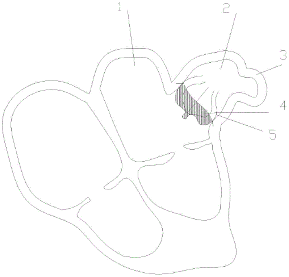
In clinical practice, this device has also exposed some problems, such as the distal end of the outer sheath has a tapered closure, and if the puncture force is not well controlled, it is easy to puncture the tip of the left atrial appendage; the knotting position of the ligature on the left atrial appendage is not easy Control, when knotting is affected by body fluids and the elasticity of the left atrial appendage itself, knotting is prone to failure; under the impact of long-term blood flow and heart beating, it is easy to untie automatically and restore the left atrial appendage to its original state

Method used


Image

  • Device for left auricle ligation and using method of device
  • Device for left auricle ligation and using method of device
  • Device for left auricle ligation and using method of device

Examples


no. 1 approach

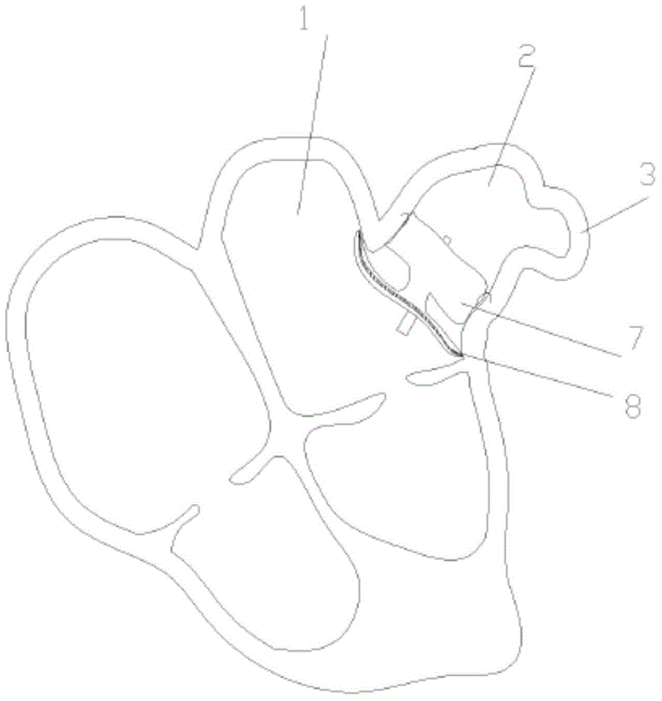
[0095] The structure of the device for left atrial appendage occlusion surgery provided by this embodiment is as follows: Figure 6-13 and Figure 16 As shown, it includes an outer sheath tube 21 for reaching the left atrial appendage through the venous blood vessel; an inner sheath tube 22 for reaching the left atrial appendage through the venous blood vessel and positioning the guide wire 23; the front end for introducing the minimally invasive surgical instrument is J A guide wire 23 of the type; a spiral device 24 of medical stainless steel material used to hold the tip of the left atrial appendage; a knotting device and a suturing device for knotting the tip of the left atrial appendage, and the suturing device can be selected for use. A suture device for minimally invasive surgery, so not shown in the figure; a cutting device for cutting off the knot at the tip of the left atrial appendage, such as thermal shears 25; wherein, the diameter of the outer sheath tube 21 is g...

no. 2 approach

[0118] In this embodiment, a sheath tube assembly for a left atrial appendage ligation operation device is provided, such as Figure 15 As shown, it includes an inner sheath 22' and the inner sheath is embedded on the inner wall of the outer sheath. The inner wall of the inner sheath tube 22' is provided with three slideways 221' for positioning the three guide wires. The three slideways 221' are arranged in an equilateral triangle on the inner wall of the inner sheath tube 22'. The inner sheath of this structure can penetrate three guide wires at the same time, and each guide wire is loaded with different surgical instruments, and the next step can be directly performed after the previous step of the operation is completed, without having to end each step of the operation. After the guide wire is replaced, the operation time is saved and the operation risk is greatly reduced.

no. 3 approach

[0120] This embodiment provides a surgical device for left atrial appendage ligation, which includes: a sheath assembly; a guide wire with a J-shaped front end for introducing minimally invasive surgical instruments; a medical stainless steel material for holding the tip of the left atrial appendage The screw device; the suturing device for knotting the tip of the left atrial appendage; the cutting device for cutting at the knot at the tip of the left atrial appendage.

[0121] The method of use of this embodiment is:

[0122] a. Find the vein entrance;

[0123] b. Puncture through the interatrial septal approach with the outer sheath so that the front end of the outer sheath touches the inside of the tip of the left atrial appendage;

[0124] c. Under the guidance of fluoroscopy and intracardiac echocardiography, insert the three guide wires into the tip of the left atrial appendage along the inner sheath;

[0125] d. Install a screw device, a suturing device and a cutting ...


PUM


Abstract

The invention discloses a device for a left auricle ligation. The device comprises an epitheca tube, an endotheca tube, guide wires, a spiral device, a knotting and sewing device and a cutting device. The epitheca tube reaches a left auricle through a vein blood vessel. The endotheca tube is used for interatrial septum puncturing and positioning the guide wires in the left auricle. The guide wires are used for being introduced into a minimally invasive surgery device. The spiral device is used for being connected with the tip of the left auricle. The knotting and sewing device is used for knotting and sewing the tip of the left auricle. The cutting device is used for cutting the knotted part of the tip of the left auricle. The caliber of the epitheca tube is larger than that of the endotheca tube. The device has the benefits that the part, prone to cause thrombus, in the left auricle is cut, so that the hidden danger of stroke is eliminated thoroughly; the guide wires are arranged in the endotheca tube to reduce the danger that the tip of the left auricle is mistakenly punctured by the epitheca tube; the whole operation is conducted in a vein blood vessel, and a thoracotomy is not needed, so that the invasive surgery is reduced, and the postoperative recovery time is shortened; and since the left auricle is cut, the problem of related complications caused by left atrial appendage occluder at present is solved.

Description

technical field [0001] The invention relates to a device for ligation of the left atrial appendage and a method for using the same, belonging to the technical field of medical devices. Background technique [0002] The atrial appendages are ear-shaped sacs that protrude from, and are part of, the atrium. In the upper part of the left atrium of the heart, it is located on the back side of the aorta and pulmonary artery, and its corner protrudes to the front right side, which is the left atrial appendage. The posterior wall of the left atrium has four venous openings, with the left atrioventricular ostium below. It is not known that the atrial appendage has any physiological function; however, because the inner surface of the left atrial appendage has a comb-like muscle and the surface is uneven, it is easy to cause the blood flow to vortex and slow down. In some pathological conditions (such as rheumatic heart disease, atrial fibrillation) , thrombus is easily formed in the...

Claims


Application Information

Patent Timeline
Patent Type & Authority Applications(China)
IPC IPC(8): A61B17/12A61B17/22
Inventor 吕斐
Owner SHANGHAI TONGJI HOSPITAL
Learn More