
Biological membrane and preparation method thereof

A biomembrane and membrane tissue technology, applied in tissue regeneration, medical science, prosthesis, etc., can solve the problem of high toxicity of polyethylene glycol octylphenyl ether, poor biocompatibility and safety, damage to collagen fiber structure, etc. problem, to achieve good decellularization and degreasing effect, improve decellularization efficiency, and less damage to collagen fibers

Inactive Publication Date: 2016-09-28
SHAANXI RUISHENG BIOTECH
View PDF5 Cites 12 Cited by
  • Summary
  • Abstract
  • Description
  • Claims
  • Application Information

AI Technical Summary

Problems solved by technology

[0003] The existing guided tissue regeneration membranes are mainly obtained through two methods. One method is artificial synthesis, which prepares a dense layer and The loose layer, or compounding hyaluronic acid, calcium salt, etc. on foreign degradable materials, is integrated into a two-layer or three-layer structure guided tissue regeneration film through cross-linking or electrospinning technology, which is obtained by artificial synthesis. The guided tissue regeneration membrane needs to introduce foreign degradable materials, and the products of degradable materials are acidic after degradation, which will cause inflammation. Therefore, the biocompatibility and safety of the guided tissue regeneration membrane are poor; making the guided tissue regeneration The mechanical strength of the membrane becomes poor, and it is prone to collapse, thereby affecting the growth of new tissues; another method is to obtain a biofilm containing only extracellular matrix by decellularizing and degreasing the animal membrane tissue. Layer structure, when the biofilm is used as a guiding tissue regeneration film for periodontal tissue defects, no foreign degradable materials are introduced, and the biocompatibility, safety, and mechanical strength are relatively excellent; however, in the prior art, all Pancreatin, EDTA, and polyethylene glycol octylphenyl ether are mainly used for decellularization of the above-mentioned animal membrane tissues. The decellularization effect of these reagents is not obvious and the efficiency is low. Enzymes have greater damage to the collagen fiber structure of the material

Method used


Image

  • Biological membrane and preparation method thereof

Examples


preparation example Construction

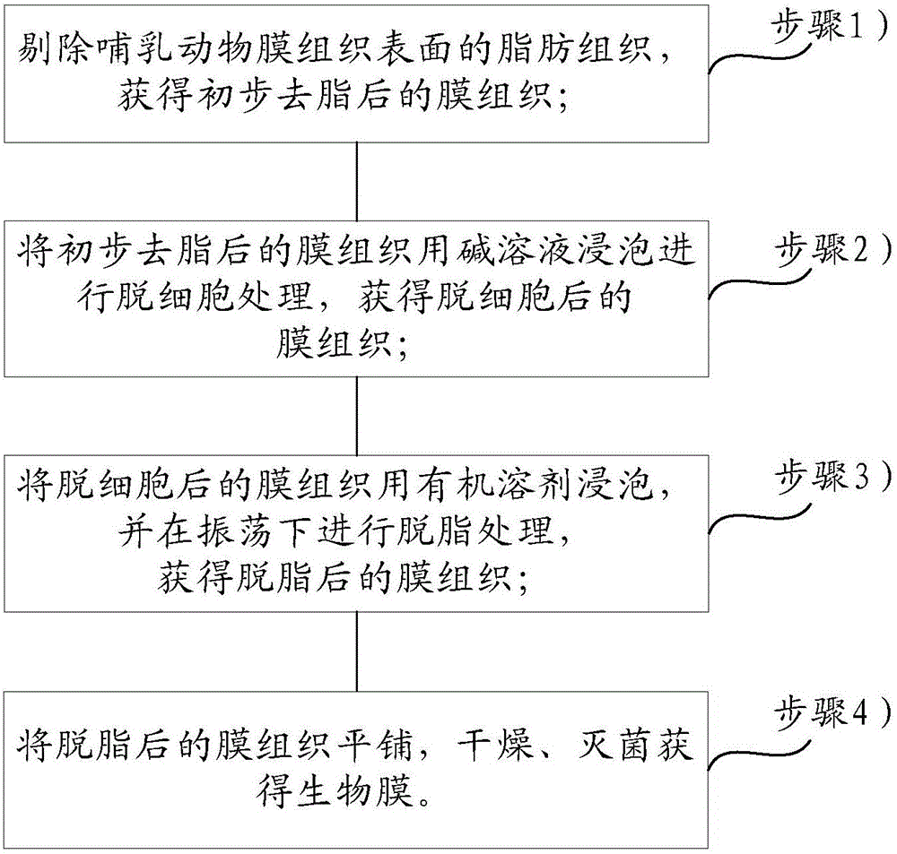
[0025] On the one hand, the embodiment of the present invention provides a kind of preparation method of biofilm, see figure 1 ,include:

[0026] Step 1) remove the fatty tissue on the surface of the mammalian membrane tissue, and obtain the membrane tissue after the preliminary fat removal;

[0027] Step 2) Soaking the membrane tissue after preliminary degreasing with alkaline solution for decellularization treatment to obtain the membrane tissue after decellularization;

[0028] Step 3) Soak the decellularized membrane tissue in an organic solvent, and perform degreasing treatment again under vibration to obtain the degreased membrane tissue;

[0029] Step 4) flattening the degreased membrane tissue, drying and sterilizing to obtain a biofilm.

[0030] Wherein, it should be noted that since the animal membranous tissue itself has a dense layer and a loose layer, the dense layer is formed by densely arranged smaller collagen fiber bundles, which can prevent soft tissue from...

Embodiment 1

[0066] S101, removing the fatty tissue on the surface of the bovine pericardium, cleaning the surface foreign matter, and obtaining the bovine pericardium after preliminary fat removal;

[0067] S102. Soak the bovine pericardium after preliminary degreasing with 5 times the amount of 1M sodium hydroxide for 1.0 h to obtain decellularized bovine pericardium;

[0068] S103. Wash the decellularized bovine pericardium for 15 minutes with 3 parts of phosphate buffer solution whose mass is 15 times that of the decellularized membrane tissue, so that the pH value of the bovine pericardium is 6.0;

[0069] S104, degreasing the bovine pericardium obtained in step S103 with 5 times the amount of isopropanol for 3 hours to obtain degreased bovine pericardium, the fat content of the defatted bovine pericardium is 1.5%;

[0070] S105. Wash the degreased bovine pericardium with 5 parts of water whose mass is 15 times that of the degreased bovine pericardium respectively, so that the mass co...

Embodiment 2

[0075] S101, removing the fat tissue on the surface of the porcine small intestine mucosa, cleaning the foreign matter on the surface, and obtaining the porcine small intestine mucosa after the initial fat removal;

[0076] S102. Soak the pig small intestinal mucosa after preliminary degreasing with 5 times the amount of 0.25M sodium hydroxide for 0.5h to obtain decellularized pig small intestinal mucosa;

[0077] S103. Wash the decellularized porcine small intestinal mucosa for 10 minutes with 3 parts of phosphate buffer solution whose mass is 15 times that of the decellularized membrane tissue, so that the pH value of the porcine small intestinal mucosa is 8.0;

[0078] S104, degreasing the porcine small intestinal mucosa obtained in step S103 with 5 times the amount of isopropanol for 1.5 hours to obtain defatted porcine small intestinal mucosa, the fat content of the defatted porcine small intestinal mucosa is 1.2%;

[0079] S105. Wash the degreased porcine small intestina...


PUM


Abstract

The invention relates to the technical field of tissue engineering, in particular to a biofilm and a preparation method thereof. This method has simple process, low cost, less damage to the collagen fibers of membrane tissue, and good decellularization effect. The obtained biofilm retains the natural double-layer structure of animal membrane tissue and can guide tissue regeneration. The clinical application of the membrane has created conditions. The embodiment of the present invention provides a method for preparing a biofilm, comprising: step 1) removing the fatty tissue on the surface of the mammalian membrane tissue to obtain the membrane tissue after the preliminary degreasing; step 2) treating the membrane tissue after the preliminary degreasing with alkali solution soaking for decellularization to obtain decellularized membrane tissue; step 3) soaking the decellularized membrane tissue in an organic solvent, and performing degreasing treatment under vibration to obtain degreased membrane tissue; step 4) degreasing The final membrane tissue was flattened, dried, and sterilized to obtain a biofilm.

Description

technical field [0001] The invention relates to the technical field of guided tissue regeneration, in particular to a biofilm and a preparation method thereof. Background technique [0002] Guided tissue regeneration is an ideal technique to promote periodontal new attachment; this technique is to cover the periodontal tissue defect with a guided tissue regeneration film to prevent fast-growing epithelial cells and fibroblasts from growing into the defect area. Thereby providing a certain space for the growth of periodontal ligament cells and bone cells and promoting the regeneration of periodontal tissue. Therefore, an ideal guided tissue regeneration membrane should have a double-layer structure with a dense layer and a loose layer, the dense layer can block the invasion of soft tissues, and the porous structure of the loose layer is more conducive to the attachment and growth of bone cells. [0003] The existing guided tissue regeneration membranes are mainly obtained th...

Claims


Application Information

Patent Timeline
IPC IPC(8): A61L27/36A61L27/50
CPCA61L27/3604A61L27/3687A61L27/50A61L2430/12A61L2430/40
Inventor 刘芮廷汪亚柱马波郜香黎
Owner SHAANXI RUISHENG BIOTECH
Learn More