HPV16E7 monoclonal antibody and preparation method and application thereof
A HPV16E7, monoclonal antibody technology, applied in the direction of antibodies, chemical instruments and methods, anti-tumor drugs, etc., can solve the problems of high false-positive rate of monitoring results and difficult implementation in small hospitals, and achieve high application value.
- Summary
- Abstract
- Description
- Claims
- Application Information
AI Technical Summary
Problems solved by technology
Method used
Image
Examples
preparation example Construction
[0035] The present invention provides a preparation method of HPV16E7 monoclonal antibody, comprising the following steps,
[0036] Step 1, HPV16E7 gene amplification
[0037] Using HPV16 positive secretion as a template for PCR amplification, the upstream primer sequence is shown in SEQNO.1, and the downstream primer sequence is shown in SEQNO.2;
[0038] Step 2, obtaining HPV16E7 recombinant protein
[0039] Using the PCR product obtained in recovery step 1 to carry out HPV16E7 expression vector construction and induced expression, and carry out the purification of HPV16E7 recombinant protein;
[0040] Step three, immunization
[0041] The antigen used is the purified HPV16E7 recombinant protein obtained in step 2;
[0042] Step 4, cell fusion to obtain hybridoma cells;
[0043] Step 5, screening positive clones of the hybridoma cells obtained in step 4 to obtain specific HPV16E7 monoclonal antibody hybridoma cells;
[0044] Step six, using the specific HPV16E7 monoclonal...
Embodiment 1
[0046] The preparation of embodiment one HPV16E7 antigen
[0047] First, design and synthesize HPV16E7 primers, and design a pair of specific primers based on the HPV16E7 gene sequence released by NCBI. Upstream primer (SEQNO.1): 5'-CGC GGATCC ATGCATGGAGATACACCTACATTG' (the underlined part is the BamHI restriction site); downstream primer (SEQNO.2): 5'-CCG CTCGAG CTATTATGGTTTCTGAGAACAGATG-3' (the underlined part is the XhoI restriction site).
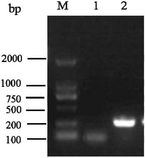
[0048] Then, the HPV16E7 gene was amplified, and the HPV16 positive secretion was used as a template for PCR amplification. The reaction system is as follows: 10×PCR buffer 2μl, MgCL 2 (25mmol / L) 1.5ul, dNTP0.4μl, upstream and downstream primers 0.5μl, template DNA 1μl, Taq enzyme 0.2μl, deionized water 13.9μl; reaction conditions: 94°C for 5min, 94°C, 45s→56°C, 45s→72°C, 45s, 30 cycles, 72°C extension for 7min, the results are attached figure 1 As shown (Note: M: marker; 1: negative specimen; 2: HPV16 positive specimen).
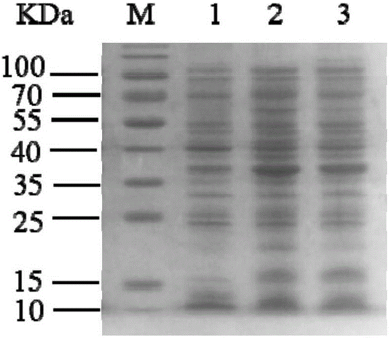
[004...
Embodiment 2
[0051] The preparation of embodiment secondary anti-HPV16E7 monoclonal antibody
[0052] 1) Immunization of mice Eight-week-old Balb / C mice were immunized with the purified HPV16E7 antigen in Example 1. The way of immunization is multi-point subcutaneous injection, the immunization dose is 0.05 mg / monkey, and the immunization interval is 2 weeks. Complete Freund's complete adjuvant was added for the first immunization, followed by three times of immunization with Freund's incomplete adjuvant, and finally the antigen solution was used as a shock immunization.
[0053] 2) Cell fusion and hybridoma cell selection After 3 days of shock immunization, mouse splenocytes were taken to fuse with SP2 / 0 cells, and HAT was used to select culture at 37°C, 5% CO 2 cultured in an incubator. After 10 days, use HPV16E7 as the antigen-coated ELISA plate (10ng / well), detect the culture supernatant and screen the positive clones by indirect ELISA, and continue to limit the dilution of the scree...
PUM
 Abstract
Description
Claims
Application Information
 - Generate Ideas
- Intellectual Property
- Life Sciences
- Materials
- Tech Scout
- Unparalleled Data Quality
- Higher Quality Content
- 60% Fewer Hallucinations
Browse by: Latest US Patents, China's latest patents, Technical Efficacy Thesaurus, Application Domain, Technology Topic, Popular Technical Reports.
© 2025 PatSnap. All rights reserved.Legal|Privacy policy|Modern Slavery Act Transparency Statement|Sitemap|About US| Contact US: help@patsnap.com


