A kind of endonuclease mus81 detection kit and its detection method and application
A detection kit and endonuclease technology, applied in the field of malignant tumor detection, can solve the problems of malignant tumors such as easy recurrence, metastasis, rapid progress, and poor prognosis, and achieve good clinical application prospects and highly targeted effects
- Summary
- Abstract
- Description
- Claims
- Application Information
AI Technical Summary
Problems solved by technology
Method used
Image

Examples
Embodiment 1
[0035] This embodiment provides a detection kit for endonuclease MUS81, including the following reagents:
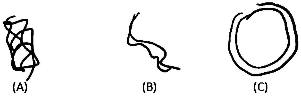
[0036] Digestion substrate: plasmid DNA.
[0037] Lysis solution: a system for lysing tumor cells, including: 0.01mol / L phosphate solution PBS (pH 7.4), 4% CHAPS, RNase inhibitor (0.2U / μl).
[0038] Digestion solution: a medium for the determination of endonuclease MUS81 activity, including: 0.01mol / L phosphate solution PBS (pH 7.4), 4% CHAPS, RNase inhibitor (0.2U / μl), 1mmol / L MgCl 2 , 0.1mmol / L DTT.
Embodiment 2
[0040] (1) Sample preparation:
[0041] 1. Collect cells: (1) Tumor tissue specimens: place the tissue in 1ml 1×PBS solution, crush the tissue with a high-speed homogenizer, filter it with an autoclaved copper wire mesh, and wash it with PBS to obtain a cell suspension ; (2) Peripheral blood samples: remove plasma, add 1:1 normal saline (NS) to the blood cell pellet according to the volume ratio, after mixing, gently add it to the ficoll separation solution (volume ratio 1:1), 1500rpm Centrifuge for 20 minutes, collect the white blood cell layer, add NS according to the volume ratio (1:4), mix well, and centrifuge at 1800rpm for 10min; remove the lower layer, add NS according to the volume ratio (1:4), mix well, and centrifuge at 1800rpm for 10min, collect a single nucleus cell. Then, use erythrocyte lysate (8.29g NH4CL+1g KHCO 3 +200ul 0.5M EDTA dissolved in 1L distilled water) to remove the red blood cells in the collected cells. (3) Isolation of purely cultured primary t...
Embodiment 3
[0054] (1) Reagent preparation:
[0055] LB solid medium containing Amp: tryptone 10g + yeast extract (yeast extract) 5g + sodium chloride 10g, add distilled water to 1L. The pH was adjusted to 7.0 with 1M NaOH, and finally autoclaved. Take the above-prepared LB culture solution and add it to agar (15g / L), after autoclaving, cool to about 60°C, add ampicillin (100mg / L) to make the final concentration 50ug / ml, shake well and spread the plate; Prepare LB plates containing ampicillin.
[0056] (2) Sample preparation:
[0057] 1. Collect cells: (1) Tumor tissue specimens: place the tissue in 1ml 1×PBS solution, crush the tissue with a high-speed homogenizer, filter it with an autoclaved copper wire mesh, and wash it with PBS to obtain a cell suspension ; (2) Peripheral blood samples: remove the plasma, add 1:1 normal saline (NS) to the blood cell pellet according to the volume ratio and mix well, then gently add it to the ficoll separation solution (1:1 volume ratio), and centr...
PUM
 Abstract
Description
Claims
Application Information
 - R&D
- Intellectual Property
- Life Sciences
- Materials
- Tech Scout
- Unparalleled Data Quality
- Higher Quality Content
- 60% Fewer Hallucinations
Browse by: Latest US Patents, China's latest patents, Technical Efficacy Thesaurus, Application Domain, Technology Topic, Popular Technical Reports.
© 2025 PatSnap. All rights reserved.Legal|Privacy policy|Modern Slavery Act Transparency Statement|Sitemap|About US| Contact US: help@patsnap.com