Culture medium and method for inducing differentiation of umbilical cord mesenchymal stem cells to corneal epithelial cells
A technology of corneal epithelial cells and amniotic membrane epithelial cells, which is applied in the field of stem cell culture, can solve problems such as differentiation difficulties, and achieve the effects of reducing risks, increasing percentages, and having a wide range of sources
- Summary
- Abstract
- Description
- Claims
- Application Information
AI Technical Summary
Problems solved by technology
Method used
Image


Examples
Embodiment 2
[0062] Example 2 Preparation of Amnion Epithelial Cells
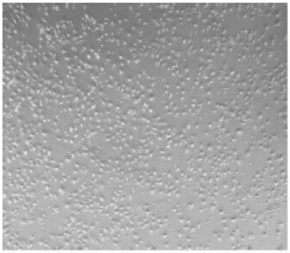
[0063] Human amniotic membrane was taken from healthy puerpera (serological examinations such as hepatitis B, hepatitis C, HIV, mycoplasma, and syphilis were all negative), and the amniotic membrane was rinsed twice in PBS containing 100 U / mL penicillin and 100 U / mL streptomycin. Cut into about 30-50cm 2 For large or small tissue pieces, add a sufficient amount of 0.25% trypsin digestion solution, digest in a 200R constant temperature oscillator at 37°C for 15-30min, take out every 10min and shake vigorously by hand, centrifuge at 2000rpm for 5min, and then add enough 2.5g / L collagenase Ⅱ+0.05g / LDnas enzyme, digest at 37°C for 4h, centrifuge, then add keratinocyte serum-free medium (Keratinocyteserum-freemedium, KFSM), 10-15ng / ml epidermal growth factor (EGF) culture solution, Beat evenly, with 1×10 7 cells / L were inoculated in a culture dish with a diameter of 10 cm, placed in a cell culture box with a volume fractio...
Embodiment 3
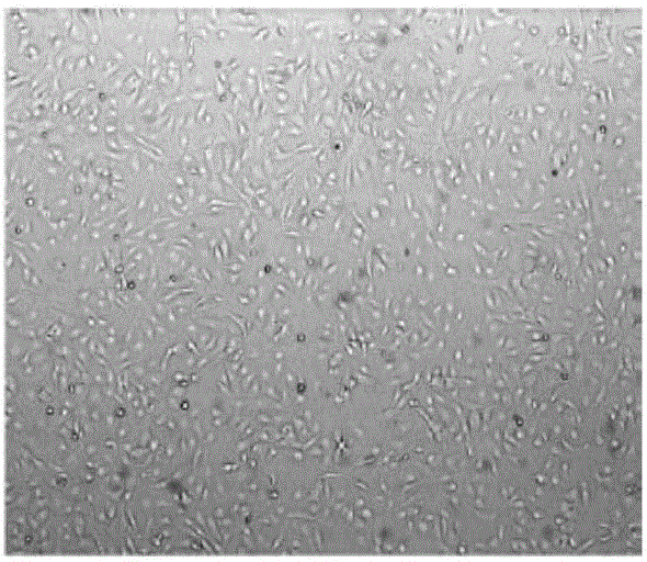
[0066] Take the 3rd-5th generation hUC-MSCs and the 2nd generation amniotic epithelial cells in the logarithmic phase of proliferation, inoculate them in the Transwell co-culture system with polylysine-treated sterile coverslips, and implant the amniotic membrane in the upper chamber Epithelial cells were seeded at a density of 4-6 x 10 5 / well, the lower chamber was implanted with hUC-MSCs, the seeding density was 1-1.5×10 5 / hole. Add 2.5ml containing 100U / mL penicillin and 100U / mL streptomycin to each well, the volume fraction is 55% LONZA human stem cell serum-free medium (LonzaUltraCULTURE TM ), 45% keratinocyte serum-free medium (Keratinocyteserum-freemedium, KFSM) culture solution, 5ng / ml epidermal growth factor (EGF), 5ug / ml insulin complete culture solution. Replace with new culture medium every 2-3 days. After 7 days of culture, the cell slides were taken out, and the immunohistochemical staining of cytokeratin AE1 and AE5 was carried out according to the instruct...
Embodiment 4
[0068] The 3rd-5th generation hUC-MSCs and the 2nd generation amniotic epithelial cells in the logarithmic phase of proliferation were inoculated in the Transwell co-culture system with polylysine-treated sterile coverslips, and the upper chamber was implanted with amniotic membrane Epithelial cells were seeded at a density of 4-6 x 10 5 / well, the lower chamber was implanted with hUC-MSCs, the seeding density was 1-1.5×10 5 / hole. Add 2.5ml containing 100U / mL penicillin and 100U / mL streptomycin to each well, the volume fraction is 55% LONZA human stem cell serum-free medium (LonzaUltraCULTURE TM ), 45% keratinocyte serum-free medium (Keratinocyteserum-freemedium, KFSM) culture solution, 10ng / ml epidermal growth factor (EGF), 10ug / ml insulin complete culture solution. Replace with new culture medium every 2-3 days. After 7 days of culture, the cell slides were taken out, and the immunohistochemical staining of cytokeratin AE1 and AE5 was carried out according to the instruc...
PUM
 Abstract
Description
Claims
Application Information
 - R&D
- Intellectual Property
- Life Sciences
- Materials
- Tech Scout
- Unparalleled Data Quality
- Higher Quality Content
- 60% Fewer Hallucinations
Browse by: Latest US Patents, China's latest patents, Technical Efficacy Thesaurus, Application Domain, Technology Topic, Popular Technical Reports.
© 2025 PatSnap. All rights reserved.Legal|Privacy policy|Modern Slavery Act Transparency Statement|Sitemap|About US| Contact US: help@patsnap.com