In-vitro glial scar forming model and establishment method and application thereof

A glial scar and construction method technology, which is applied in the field of cell biology, can solve the problems of enhancement, no successful in vitro glial scar formation model, unsatisfactory supply and price, etc., and achieves the effect of simple preparation.

Active Publication Date: 2015-09-16
南通大学技术转移中心有限公司
View PDF1 Cites 3 Cited by
  • Summary
  • Abstract
  • Description
  • Claims
  • Application Information

AI Technical Summary

Problems solved by technology

So far, there are several culture models of glial scars as follows: (1) Use nitrocellulose sheets to insert into the cortex, then remove them, and culture them on the tissue adhered to the surface (Rudge JS, Silver J. Inhibition of neurite outgrowth on astroglial scars in vitro.J Neurosci.1990; 10:3594-3603.); although this method can obtain the corresponding components of glial scars in vitro, it is not a structure formed by in vitro cell culture, so the entire glial scars form process cannot be observed
(2) Using astrocytes and brain (spinal) membrane fibroblasts to directly co-culture to produce substances and related effects similar to glial-limiting membrane (Ness R, David S. Leptomeningeal cells modulate the neurite growth promoting properties of astrocytes in vitro. Glia. 1997; 19:47-57. Struckhoff G. Cocultures of meningeal and astrocytic cells–a model for the formation of the glial-limiting membrane. Int J Dev Neurosci. 1995; 13:595 -606.); This method can observe the activation process of astrocytes, but in the process of simulating the formation of glial boundary membrane, it may be closer to the process of normal development of nervous tissue, and there is no correlation when the central nervous system is damaged factors involved
(3) Use 3D glue to solidify the inhibitory proteoglycan and other substances of glial scars, and observe the effect on the growth of neurons and other cells (Gilbert RJ, McKeon RJ, Darr A, Calabro A, Hascall VC, Bellamkonda RV.CS-4, 6is differentially upregulated in glial scar and is a potent inhibitor of neurite extension. Mol Cell Neurosci.2005; 29(4):545-58.); but this method does not involve cells forming glial scar, so it is only for a certain To study the effect of an inhibitory substance
(4) Culture astrocytes and cerebrospinal fibroblasts on a silicone rubber petri dish, and then form cell wounds through appropriate mechanical stretching, which can make astrocytes appear star-shaped and aggregate into clusters , and the marker expression of glial scar was significantly enhanced (Wanner IB, Deik A, Torres M, Rosendahl A, Neary JT, Lemmon VP, Bixby JL.A new in vitro model of the glial scar inhibits axon growth.Glia.2008; 56:1691-709.); This method requires special silicone rubber cell culture dishes, and its supply and price are not satisfactory
(5) Utilizing the action of β-transforming growth factor, glial cells and cerebrospinal fibroblasts aggregate into clusters and the characteristic markers of glial scars appear (Kimura-Kuroda J, Teng X, Komuta Y, Yoshioka N, Sango K, Kawamura K, Raisman G, Kawano H. An in vitro model of the inhibition of axon growth in the lesion scar formed after central nervous system injury. Mol Cell Neurosci. 2010; 43:177-87.); of course This method is the result of the direct action of bioactive molecules, but when the central nervous system is injured, the change in the level of bioactive molecules is not the first factor in the formation of glial scars, but the secondary result. Glial scar formation is then induced; therefore, this method cannot fully reproduce the entire process of glial scar formation
[0006] From the above-mentioned experimental models of glial scars that have appeared in vitro, it can be seen that there are still many shortcomings. At present, an ideal model of glial scar formation in vitro has not been successfully constructed.

Method used


Image

  • In-vitro glial scar forming model and establishment method and application thereof
  • In-vitro glial scar forming model and establishment method and application thereof
  • In-vitro glial scar forming model and establishment method and application thereof

Examples


Embodiment 1

[0065] Example 1: Cultivation, purification and identification of brain (spinal) membrane fibroblasts and astrocytes

[0066] 1. Cultivation, purification and identification of brain (spinal) membrane fibroblasts

[0067] 1.1 Isolation of the brain

[0068] SD rats 24 hours after birth were soaked in 75% alcohol for disinfection; the ophthalmic curved scissors were used to decapitate the head, and the head was fixed with the thumb and forefinger, and the skull skin was cut with ophthalmic curved scissors; The skull was cut open and turned to both sides with the “people” seam. At this time, the cerebral hemispheres on both sides were exposed. The whole brain was separated from the base of the skull using curved micro-tweezers, and placed in pre-cooled HBSS buffer.

[0069] 1.2 Isolation of cerebral meninges and cerebrospinal fibroblasts for culture and purification

[0070] Under a stereomicroscope, the cerebral cortex is facing upwards, and the brain is fixed with one micros...

Embodiment 2

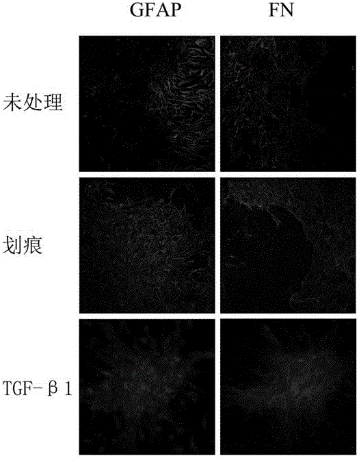
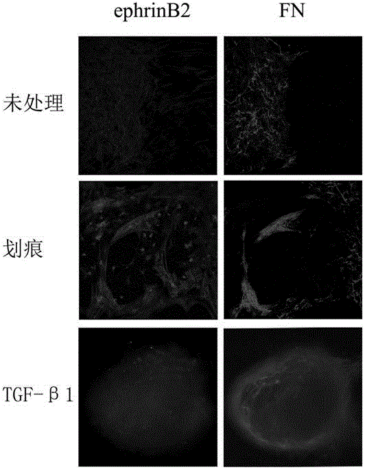
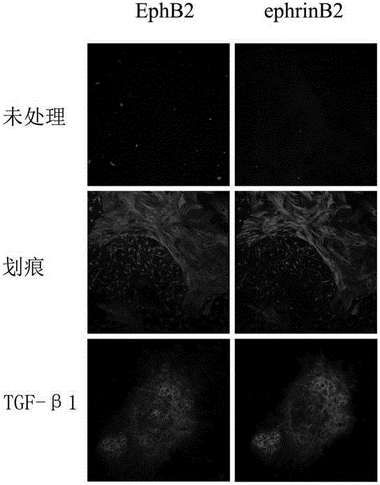
[0082] Example 2: Establishment and identification of the glial-like scar model of the present invention

[0083] 3.1 Establishment of glial-like scars

[0084] Purified 6×10 4 cerebrospinal fibroblasts and 8×10 4 Astrocytes were respectively planted in adjacent chamber slides (Nunc Company) chambers, the slides at the bottom of the chambers had been pre-coated with 50 μg / ml L-polylysine, about 200 μl per chamber Cell suspension (the density ratio of brain (spinal) membrane fibroblasts and astrocytes is 3:4), remove the small chamber after 0.5h, add 5ml DMEM / F12 medium containing 10% FBS, in 37°C, 5% CO 2 Cultivate in an incubator for about 3-5 days. The two types of cells have moved closer to each other. Take the needle tip of a disposable 1ml syringe. Under the microscope, there are scratches in the word "Feng" at the junction of the two cells, including 16 vertical scratches and 1 horizontal scratch. Change the medium. Afterwards, the culture was continued, and a glial-...


PUM


Abstract

The invention belongs to the technical field of cytobiology and provides an establishment method of an in-vitro glial scar forming model. The establishment method of the in-vitro glial scar forming model comprises the steps that purified meninx (meningo) fiber cells and purified astroglia cells are planted in adjacent small cells on a cavity glass slide respectively with a certain amount ratio, and a culture medium is added for culture; after the cells grow in an adherent mode, a partition board between the small cells is removed, and culture continues to be conducted; when it is observed that the meninx (meningo) fiber cells and the astroglia cells grow and come close to each other, mechanical scratching is conducted at the junction of the two kinds of cells so as to damage the cells, and finally, a glial scar structure is formed. The in-vitro glial scar forming model obtained through the method can well simulate the whole forming process of an in-vitro glial scar, the corresponding glial scar form structure and the change relevant to the cytobiology and the molecular biology appear, and an in-vitro cell-level experimental model is provided for studying of the glial scar forming mechanism and relevant treatment.

Description

technical field [0001] The invention belongs to the technical field of cell biology, and specifically relates to a model that can simulate the formation of glial scars after central nervous system injury in vitro and its construction method, and provides in vitro cellular level for research on the mechanism of glial scar formation and related treatment research. experimental model. Background technique [0002] When the central nervous system is injured, it will cause the quiescent astrocytes to transform into reactive astrocytes, and protrude star-shaped thick processes around the injury site; at the same time, the brain (spinal) membrane fiber cells will also migrate to the injury site. , distributed between the injured tissue and reactive astrocytes, arranged tightly; reactive astrocytes will extend finger-like processes to the brain (spinal) membrane fibroblasts, and combine with each other; the two types of cells secrete together And form a special extracellular matrix...

Claims


Application Information

Patent Timeline
IPC IPC(8): C12N5/071C12N5/079C12Q1/02
Inventor 王晓冬李奕谈玲陈雪林巍巍陈颖潘静莹陆冰缪玲玉
Owner 南通大学技术转移中心有限公司
Why Eureka
Try Eureka