
Sample preparation method for mice brain tissue proteome analysis

A proteome and sample preparation technology, applied in the field of proteomics research, can solve the problems of isoelectric focusing failure, protein loss, protein spot reduction, etc., to achieve the effect of ensuring purity and concentration, increasing solubility, and improving efficiency

Active Publication Date: 2015-09-02
INST OF MODERN PHYSICS CHINESE ACADEMY OF SCI
View PDF3 Cites 5 Cited by
  • Summary
  • Abstract
  • Description
  • Claims
  • Application Information

AI Technical Summary

Problems solved by technology

In the process of protein sample preparation, kits for removing albumin and immunoglobulin IgG in blood can also be used, but the kits are expensive, the sample volume that can be processed is small, and protein loss will occur during processing
[0008] (2) There is no step of heating the protein sample to increase the solubility, and the protein extraction efficiency is not good
However, the temperature of the protein lysate containing urea should not exceed 37°C, otherwise the urea will be hydrolyzed into isocyanate, and the isocyanate will modify the protein through carbamylation, resulting in artificial "spot series", affecting the separation and identification of protein samples
Therefore, according to this method, the sample cannot be heat-treated to solubilize
[0010] (3) There is no special step to remove lipids in brain tissue, resulting in lipids affecting protein separation and reducing protein spots
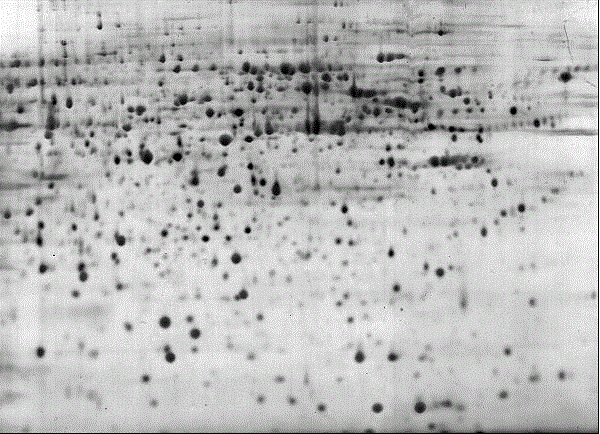
[0011] Brain tissue contains a lot of lipids. If the lipids are not removed, the viscous lipids will block the pores of the gel during proteome analysis, preventing the entry of proteins, resulting in fewer protein spots and a darker gel background. good effect
[0012] (4) There is no step to remove salt ions, which affects the electrophoresis voltage
Lower voltage will prolong isoelectric focusing time, cause isoelectric focusing failure or reduce protein separation efficiency

Method used


Image

  • Sample preparation method for mice brain tissue proteome analysis

Examples


Embodiment Construction

[0041] A sample preparation method for proteome analysis of mouse brain tissue, comprising the following steps:

[0042] (1) Apical perfusion blood replacement: take a mouse, fix the limbs, and inject chloral hydrate with a mass concentration of 10% intraperitoneally according to 0.03ml / 10g~0.05ml / 10g; The septum was removed to expose the heart; then the needle was inserted into the left ventricle, the right atrial appendage was cut, the peristaltic pump was turned on, and the apical perfusion was performed slowly with normal saline, and the brain was stopped when the liver turned grayish white.

[0043] (2) Protein extraction: Put the brain tissue obtained in step (1) into a mortar filled with liquid nitrogen and grind until the brain tissue is ground into powder; then weigh 0.2~0.5g of brain tissue powder and transfer it to a 1.5ml EP centrifuge tube , according to the ratio of 300μl: 0.1g, add 4 ℃ pre-cooled pH=8.1, 50mM Tris buffer to the EP centrifuge tube; secondly, put ...


PUM


Abstract

The invention relates to a sample preparation method for mice brain tissue proteome analysis. The method comprises the following steps: (1) replacing the blood through apex perfusion, namely, narcotizing a mice, performing slow apex perfusion, stopping while the liver is in grey white, and taking out a brain tissue; (2) extracting protein, namely, grinding the brain tissue into powder, adding a pre-cooled Tris buffer solution, ultrasonically treating, incubating in a shaking table, and centrifuging to obtain a protein extracting solution I; (3) thermally treating to increase protein solubility, namely, mixing the protein extracting solution I and the thermally treated solution, treating for 5min, and centrifuging to obtain the protein extracting solution II; (4) precipitating to remove fat, namely, adding the protein extracting solution II in the pre-cooled fat-removing precipitation solution CUS, precipitating, centrifuging, washing, and drying to obtain purified protein powder; and (5) re-dissolving the protein, namely, adding the purified protein powder in an urea protein lysate, and centrifuging to obtain a proteome sample. With the sample preparation method disclosed by the invention, blood and fat influences of the brain tissue of the mice can be economically and efficiently removed, and the solubility of the protein sample is increased.

Description

technical field [0001] The invention relates to the field of proteomics research, in particular to a sample preparation method for mouse brain tissue proteome analysis. Background technique [0002] Life science research has entered the omics era of overall life research. With the development of genomics research, researchers have found that genes, as the carrier of genetic information, can only determine the basic form of life, and the genome knowledge obtained now cannot tell us The role of genes in living organisms and how they function, and the product protein expressed by genes is the real executor of life functions. Therefore, in order to understand genes and functions in depth, it is necessary to interpret and interpret at the protein level. However, there is no strict linear relationship between gene and protein expression. The expression of protein has the particularity of time and space, and its complexity and quantity are much higher than those of genes. The comp...

Claims


Application Information

Patent Timeline
IPC IPC(8): G01N33/48
Inventor 吴庆丰李强叶飞金晓东
Owner INST OF MODERN PHYSICS CHINESE ACADEMY OF SCI
Learn More