
Application of method for degrading blood to obtain monosaccharide and detecting monosaccharide in cancer detection

A cancer detection and monosaccharide technology, applied in the field of medicine, can solve the problems of low sensitivity of early cancer patients, difficulty in blood biomarkers, and large amount of data processing, etc., and achieve the effect of easy popularization of the method, short detection time and low detection cost

Active Publication Date: 2014-08-06
OCEAN UNIV OF CHINA
View PDF1 Cites 8 Cited by
  • Summary
  • Abstract
  • Description
  • Claims
  • Application Information

AI Technical Summary

Problems solved by technology

However, the sensitivity of early cancer patients is much lower than that of advanced cancer patients, which may limit the application of this method in the early diagnosis of cancer
In general, omics analysis is more comprehensive than the current cancer biomarkers, but it is generally complex, expensive, and requires a large amount of data processing, and it is not yet simple, fast, stable, reliable, and cheap clinical requirements
[0005] At present, there have been many reports of blood sugar biomarkers for cancer. However, using the current multi-step separation and identification technology to find blood biological markers for early diagnosis of cancer with sensitivity and selectivity that meet clinical requirements from more than 7,000 sugar structures. Markers are very difficult, and it is difficult to have clinical application value in the short term

Method used


Image

  • Application of method for degrading blood to obtain monosaccharide and detecting monosaccharide in cancer detection
  • Application of method for degrading blood to obtain monosaccharide and detecting monosaccharide in cancer detection

Examples


Embodiment 1

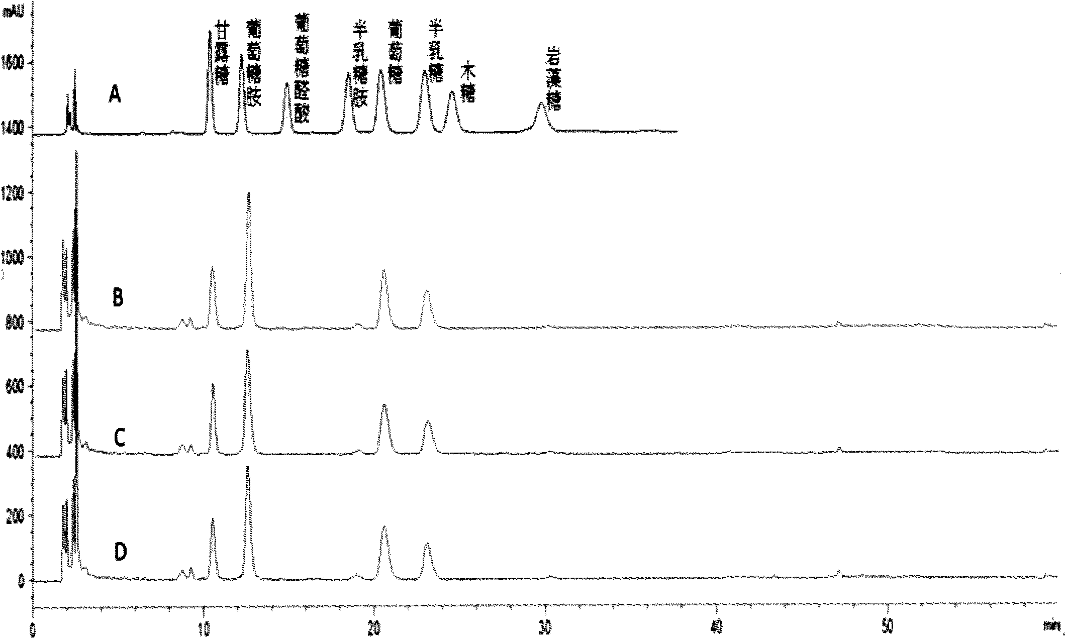
[0032] (1) Prepare solutions with different concentrations (0.5mg / mL, 0.25mg / mL, 0.1mg / mL, 0.05mg / mL, 0.025mg / mL, 0.01mg / mL) containing 8 kinds of monosaccharide standards, and then add NH 3 ·H 2 O to adjust the pH to 8-14, mix well and let stand at room temperature, then add 0.5M PMP to the water bath.

[0033] (2) Take out the reactant and cool to room temperature, add HAc to neutralize.

[0034] (3) Add chloroform to each tube for extraction, discard the lower chloroform layer, and repeat three times. The obtained water layer was passed through a 0.22um filter membrane for liquid phase analysis.

[0035] Chromatographic conditions

[0036] Chromatographic column: ZORBAX SB-C18 (5um, 4.6mm×150mm), 0.1M phosphate (13.6gKH 2 PO 4 , 1.8gNaOH) (pH6.7) buffer-acetonitrile (volume ratio 83:17) isocratic elution, flow rate: 1.0mL / min, detection wavelength: 245nm, injection volume: 20uL.

[0037] To obtain the liquid chromatograms of 8 kinds of monosaccharide standards, see ...

Embodiment 2

[0040] (1) Take 1 mL of blood from 42 normal people and centrifuge at 3000 r / min for 5 min to separate serum or plasma.

[0041] (2) Take 100uL of plasma to the ampoule, add 1mL of 2M C to each tube 2 HF 3 o 2 .

[0042] (3) Seal the tube and put it in an oil bath at 105°C for 6 hours to obtain a hydrolyzed sample solution.

[0043] (4) Cool the hydrolyzed sample solution to room temperature, transfer to a 1.5mL centrifuge tube, and condense at -4°C for 5h to remove C 2 HF 3 o 2 .

[0044] (5) Add 100uL methanol to each tube, centrifuge and concentrate to remove residual C 2 HF 3 o 2 ,repeat three times.

[0045] (6) Each tube of the obtained dry sample is dissolved in water, and then NH 3 ·H 2O adjust the pH to 8-14, mix well and let stand at room temperature, then add 0.5MPMP to the water bath.

[0046] (7) Take out the reactant and cool to room temperature, add HAc to neutralize.

[0047] (8) Add CHCl to each tube 3 After extraction, the lower layer of CHCl w...

Embodiment 3

[0052] Get 52 routine lung cancer patient blood 1mL, all the other steps are the same as embodiment 2, embodiment 3 repeats 3 times, statistical detection result, see figure 2 .


PUM


Abstract

The invention belongs to the field of medicines, and relates to an application of a method for degrading all carbohydrate chains in the blood to be monosaccharide and detecting the monosaccharide in cancer detection. A sample in the method for degrading the blood to obtain the monosaccharide and detecting the monosaccharide is the blood, and the cancer includes lung cancer, gastric cancer, ovarian cancer, penis cancer, esophagus cancer, oral cancer, biliary duct cancer, breast cancer, adenocarcinoma perampullaire, rectal cancer and bladder cancer. The method has the characteristics of simplicity and easiness in operation steps, easiness in popularization, short detection time, low requirement on the instrument, low detection cost, less consumption of blood and the like. The result displays that based on the content of eight monosaccharides in the blood, not only can a normal person and a cancer patient be distinguished, but also different cancers can be distinguished. The method for simplifying glycomics knowledge and applying in the detection of the biomarker of blood is first created in the world.

Description

technical field [0001] The invention belongs to the field of medicine, and relates to the application of a method for degrading blood to obtain and detect monosaccharides in cancer detection. Background technique [0002] Cancer is one of the leading causes of death worldwide. Cancers accounted for 7.4 million deaths in 2004 (approximately 13% of all deaths), with lung, stomach, colon, liver and breast cancers responsible for the majority of cancer deaths each year. WHO estimates that without intervention, 84 million people will die from cancer between 2005 and 2015. Cancer is the uncontrolled growth and spread of cells. It can affect almost any part of the body. Tumors often invade surrounding tissue and can metastasize to other sites. A large proportion of cancers can be cured with surgery, radiation therapy, or chemotherapy, especially if caught early. For those early-stage lung cancer patients who are detected and treated before the cancer cells spread, the average ...

Claims


Application Information

Patent Timeline
Patent Type & Authority Applications(China)
IPC IPC(8): G01N30/02
Inventor 张丽娟曾璇兰莹邱培菊曾洋洋徐玲玲周紫婧韩章润
Owner OCEAN UNIV OF CHINA
Learn More