
A kind of preparation method of decellularized cornea

A decellularized and decellularized cornea technology, applied in the field of acellular cornea preparation, can solve the problems of tissue destruction, host rejection, poor tissue fusion effect, etc. Effect

Active Publication Date: 2015-09-09
SHAANXI BIO REGENERATIVE MEDICINE CO LTD
View PDF4 Cites 0 Cited by
  • Summary
  • Abstract
  • Description
  • Claims
  • Application Information

AI Technical Summary

Problems solved by technology

The initial decellularization method was treated with TritonX100 combined with trypsin. Although this method can completely remove the cells in the tissue, it severely damaged the collagen microstructure and glycosaminoglycan components of the natural cornea, resulting in the loss of the natural cornea after treatment. Most of the tissue characteristics of the cornea, the fusion effect with the tissue after transplantation is poor, and the therapeutic purpose cannot be achieved
In order to avoid severe damage to the corneal stroma structure, only detergents (such as TritonX100, sodium dodecylsulfonate, sodium deoxycholate, etc.) The detergent still has a great degree of damage to the tissue, and the residue of the detergent is difficult to wash off, which may easily cause the cytotoxicity of the decellularized cornea
In addition, phospholipases that specifically destroy cell membranes are used instead of broad-spectrum enzymes for decellularization treatment, but it is still difficult to completely remove cell antigens, and strong immune host reactions will be induced after transplantation; Sodium cholate) combined use, still cannot avoid the impact on the natural corneal stroma, and phospholipase is very expensive, it is difficult to realize the commercialization of decellularized cornea
Japanese scholars used low temperature and high pressure method to treat porcine cornea to achieve decellularization effect, but the high pressure method has very strict requirements on equipment, and corneal collagen will also be denatured under high pressure conditions, which will affect the therapeutic effect of decellularized cornea
[0006] In order to avoid damage to the microstructure of the decellularized cornea by chemical detergents and biological enzymes, some scholars use cross-linking methods to strengthen the corneal structure. Traditional cross-linking agents (such as glutaraldehyde, EDC, etc.) It causes the corneal stroma to lose the elasticity and toughness of the natural cornea, and more seriously, the biocompatibility of the corneal stroma decreases. After transplantation, it does not fuse with the implant bed and is eventually rejected by the host.

Method used


Image

  • A kind of preparation method of decellularized cornea
  • A kind of preparation method of decellularized cornea
  • A kind of preparation method of decellularized cornea

Examples


Embodiment 1

[0041] Example 1. Preparation of decellularized (pig) cornea

[0042] Step 1. Corneal epithelium removal: Put the porcine cornea with complete structure, no damage, and 0.3 cm wide sclera into 0.2M EDTA solution, soak for 12 hours, and then use deionized water to swell the cornea by shaking method, so that the cornea reaches 1cm thick, then rub off the corneal epithelial cell layer, exposing the corneal descemet;

[0043] Step 2. Ultraviolet crosslinking: place the cornea obtained in step 1 with the elastic layer facing upwards, and irradiate it with ultraviolet light with a wavelength of 370 nm for 1 hour at room temperature to complete the crosslinking treatment;

[0044] Step 3. Virus inactivation: use a knife to draw 10 scratches on the surface of the Descemet's membrane of the cornea, and the depth of the scratches is less than 1mm; soak the cornea in 50mg / ml trehalose solution for 3 hours, and transfer to the cornea containing trehalose concentration of Soak in a mixe...

Embodiment 2

[0049] Example 2. Preparation of decellularized (rabbit) cornea

[0050] Step 1. Corneal epithelium removal: Put the rabbit cornea with complete structure, no damage, and 1 mm wide sclera in 0.1M EDTA solution, soak for 12 hours; then use deionized water to swell the cornea by shaking method to make the cornea Reach 0.5cm thick, then rub off the corneal epithelial cell layer, exposing the corneal descemet;

[0051] Step 2. Ultraviolet cross-linking: place the cornea obtained in step 1 with the anterior elastic layer facing up, and irradiate it with ultraviolet light with a wavelength of 370 nm for 20 minutes at room temperature to complete the cross-linking treatment;

[0052] Step 3. Virus inactivation: use a knife to draw 4 scratches on the surface of the Descemet membrane of the cornea, the depth of the scratches is <1mm; soak the cornea in a trehalose solution with a concentration of 20mg / ml for 0.5h; Soak in a mixed solution with a sugar concentration of 5mg / ml and an ...


PUM

PropertyMeasurementUnit
thicknessaaaaaaaaaa
thicknessaaaaaaaaaa

Abstract

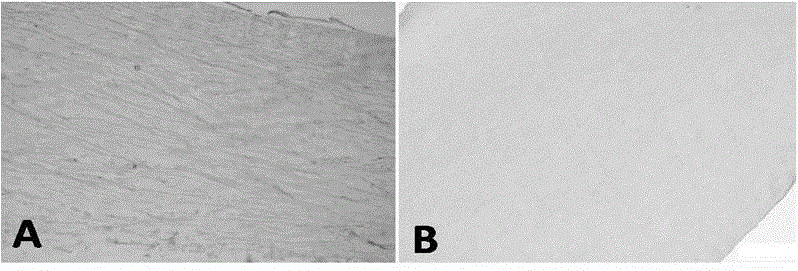
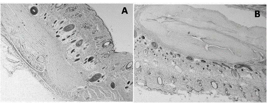
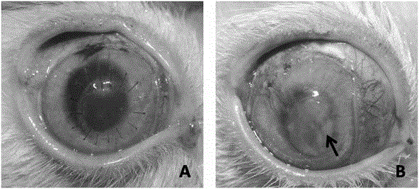
The invention relates to a decellularization cornea preparation method, which adopts steps of corneal epithelium layer removing, ultraviolet cross-linking, viral inactivation, decellularization treatment, gradient dehydration, radiation protection and sterilization. According to the present invention, the prepared decellularization cornea has characteristics of complete antigen removing, no excitation of host acquired immunity reaction, good biocompatibility, low damage on nature corneal stroma, maintaining of structure characteristics of the nature corneal, and maintaining of effective components of the nature corneal so as to provide physical and chemical properties similar to the nature corneal; and after the prepared decellularization cornea is transplanted, animal experiment results show that characteristics of transparent transplanted cornea, no scar formation, no melting generation and no neovascularization are provided, the transplanted cornea and the recipient bed are completely integrated after transplanting a month, the transplanted cornea is subjected to complete epithelization after three months, corneal stroma cells migrate toward the decellularization cornea graft so as to prove that the tissue just takes place slow reconstruction, and the transplanted cornea after 6 months does not show significant difference in histology and appearance detection compared with the nature cornea.

Description

technical field [0001] The invention belongs to the technical field of tissue engineering biomedical materials, and in particular relates to a preparation method of decellularized cornea. Background technique [0002] The cornea is one of the most important tissues to maintain normal vision in the human body. The cornea can cause vision loss or blindness due to keratoconus, bullous cornea, fungal infection, traumatic scar and other diseases. Today, corneal blindness has become the second leading cause of blindness in the world. At present, the most effective way to treat corneal blindness is allogeneic cornea transplantation. However, the lack of sources of allogeneic cornea has resulted in a large number of patients not being treated in time and eventually blind. According to statistics, more than 10 million people in the world suffer from corneal blindness, but only less than 120,000 patients can receive allogeneic cornea transplantation. [0003] In order to solve the p...

Claims


Application Information

Patent Timeline
Patent Type & Authority Patents(China)
IPC IPC(8): A61L27/36
Inventor 罗海浪
Owner SHAANXI BIO REGENERATIVE MEDICINE CO LTD
Learn More