Cell microscopic imaging method, image processing method and imaging analysis system
A microscopic imaging and analysis system technology, applied in image data processing, image enhancement, material analysis using radiation, etc., can solve the problems of ignoring influence, relying on X-ray imaging, less research in image processing, etc. and cost reduction
- Summary
- Abstract
- Description
- Claims
- Application Information
AI Technical Summary
Problems solved by technology
Method used
Image
Examples
Embodiment 1
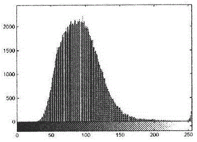
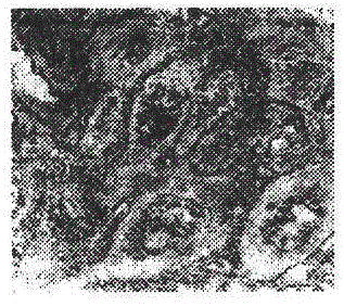
[0044] Example 1: Soft X-ray imaging and treatment of suspected esophageal squamous cell carcinoma cells using soft X-ray microscopic imaging
[0045] Select 6 hospitalized suspected patients (including 4 males and 2 females), aged 51-64 (average 57.3 years old).
[0046] The specimens were cut and sent to the pathology department for routine histological and cytological examinations. When suspected esophageal squamous cell carcinoma cells were found, light microscopic photos of the suspected esophageal squamous cell carcinoma cells were taken under an optical microscope with a magnification of 100 times. At the same time, samples of suspected esophageal squamous cell carcinoma cells were sent to the synchrotron radiation laboratory for soft X-ray microscopic beamline scanning diffraction, developed on photoresist, and soft X-ray microscopic images were taken.
[0047] The equipment used in this example is:
[0048] Use the soft X-ray microscopic imaging beamline station of t...
Embodiment 2
[0059] Example 2: Soft X-ray imaging and processing method of suspected lung cancer cells using soft X-ray microscopic imaging
[0060] Suspected lung cancer patients were selected, male or female.
[0061] Experimental means such as instruments and equipment:
[0062] Soft X-ray Microimaging Beamline Station (National Synchrotron Radiation Laboratory, University of Science and Technology of China);
[0063] Vacuum drying oven, CJ-3A glue throwing machine (produced by Beijing Semiconductor Equipment Factory);
[0064] Olympus differential interference contrast microscope (Japan)
[0065] Scanning electron microscope KYKY-1000B (manufactured by the Scientific Instrument Factory of the Chinese Academy of Sciences);
[0066] 151II ultramicrotome (manufactured by SEIXY factory in West Germany).
[0067] Preparation of soft X-ray micrograph specimens of suspected lung cancer:
[0068] Step 1: Place suspected lung cancer tissue and cell specimens on a pathological ultrathin sli...
Embodiment 3
[0081] Example 3: Application of Soft X-ray Microscopic Imaging in Plant Leaf Inspection
[0082] Take wheat seeds, germinate the seeds in an incubator at 25°C, cut the leaves on the 7th-9th day, and cut them into 2mm 2 The slice thickness is 10-20nm. The specimen is placed on the water surface of a 40°C water tank, so that the specimen floats on the water surface and spreads out quickly and evenly in a plane shape. The bath contains Tween 20 with a weight of 3ppm. The ultra-thin section specimens that have been floated and flattened in the water tank are picked up with a net, and then the electron microscope copper net is placed on absorbent paper to control the moisture and dried. The electron microscope copper net is 300 mesh and is processed as follows before the section is taken: Spread the clean copper grid on the filter paper in the petri dish, then slowly pour 0.3% by weight of Formvar solution until it covers the copper net, then drain the liquid in the petri dish imm...
PUM
| Property | Measurement | Unit |
|---|---|---|
| thickness | aaaaa | aaaaa |
| thickness | aaaaa | aaaaa |
| thickness | aaaaa | aaaaa |
Abstract
Description
Claims
Application Information
 - R&D
- Intellectual Property
- Life Sciences
- Materials
- Tech Scout
- Unparalleled Data Quality
- Higher Quality Content
- 60% Fewer Hallucinations
Browse by: Latest US Patents, China's latest patents, Technical Efficacy Thesaurus, Application Domain, Technology Topic, Popular Technical Reports.
© 2025 PatSnap. All rights reserved.Legal|Privacy policy|Modern Slavery Act Transparency Statement|Sitemap|About US| Contact US: help@patsnap.com


