
Wavelength multiplexing endoscope

a technology of endoscope and beam beam, which is applied in the field of apparatus for the illumination of endoscopic and borescopic fields, can solve the problems of reducing the risk of infection, reducing the hospital stay of patients, and typical endoscopic procedures requiring a large amount of equipment, so as to reduce the time of procedure, wide color gamut, and increase confiden

Active Publication Date: 2009-11-26
VIVID MEDICAL
View PDF20 Cites 138 Cited by
  • Summary
  • Abstract
  • Description
  • Claims
  • Application Information

AI Technical Summary

Benefits of technology

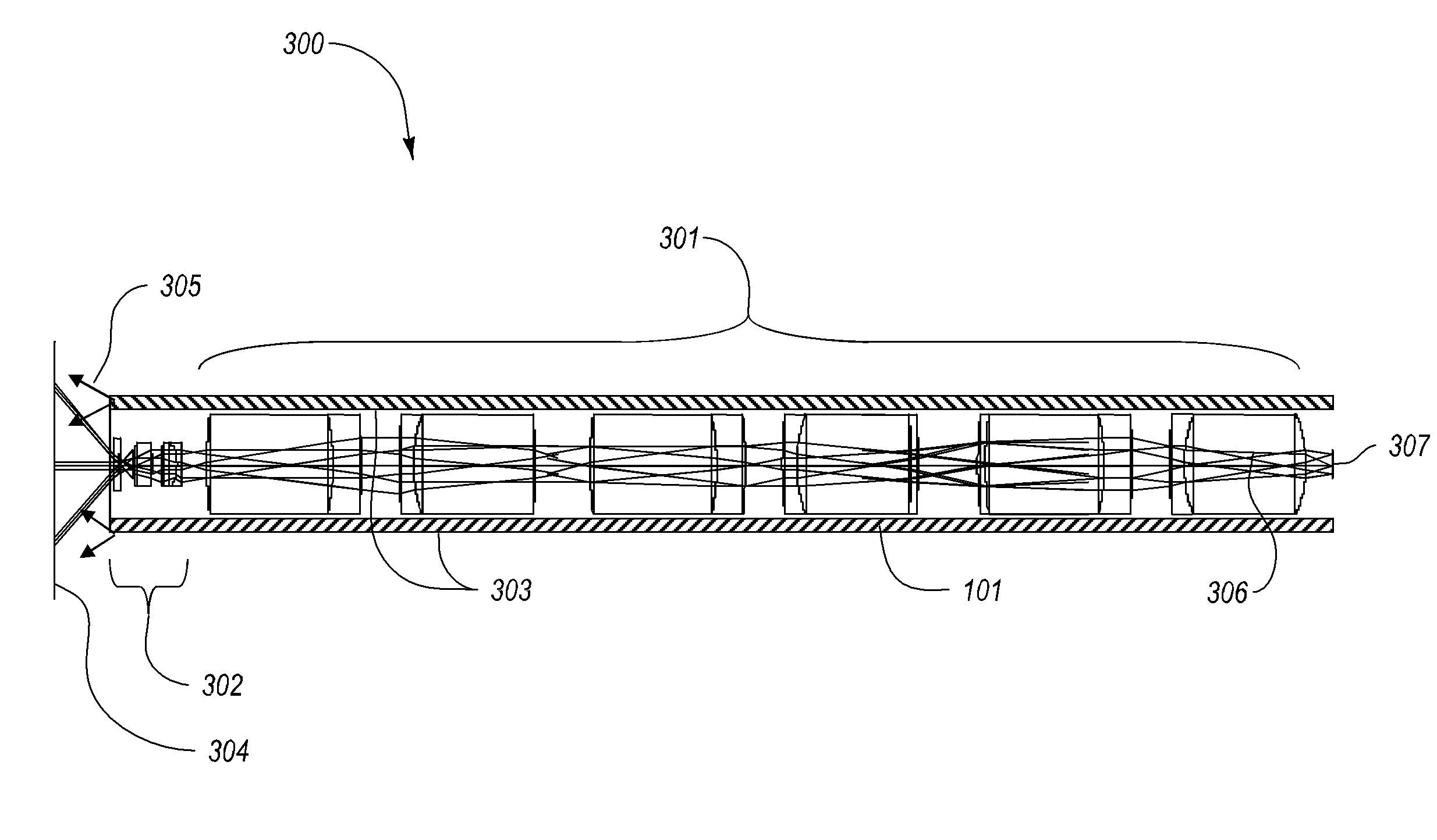
"The current disclosure describes a way to use wavelength multiplexing to capture images from multiple optical ports at the end of an endoscope using a single channel and single image sensor. This allows for stereo vision and reduces the size of the endoscope and camera module. The use of time synchronized solid state illumination and wavelength multiplexing optics can capture wide color gamut images in a single sensor flexible endoscope. A wireless embodiment is also presented where a single sensor can communicate with separate electronics and display systems to achieve stereo vision."

Problems solved by technology

This both reduces the risk of infection and reduces the patient's hospital stay.
Typical endoscopic procedures require a large amount of equipment.
However, large scope diameters are not desirable for certain operations where space is limited on the operation site.
The construction and design of this welded joint is often a weakness in the endoscope manufacturing and use, where after many operations, high temperature and high humidity sterilizations, and successive handling, this welded joint could get damaged and break, exposing the internal parts of the endoscope to the environment when the seal is broken.
Accordingly, a number of weaknesses and disadvantages exist with respect to conventional endoscopes and corresponding methods and systems.

Method used


Image

  • Wavelength multiplexing endoscope
  • Wavelength multiplexing endoscope
  • Wavelength multiplexing endoscope

Examples


Embodiment Construction

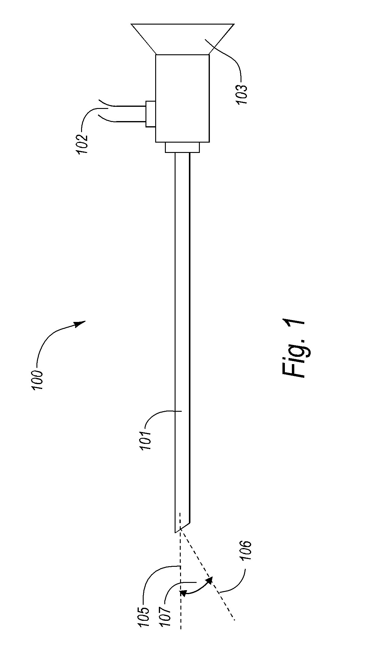
[0047]Exemplary embodiments of the disclosure concern monochromatic or polychromatic solid state light sources such as high power Light Emitting Devices (LEDs) and Laser Diodes as a means of illumination in a diagnostic or surgical endoscopic procedures, or functional borescopic systems, and how these polychromatic light sources can be used to achieve wavelength multiplexed imaging in the endoscope. In particular, these solid state light sources are incorporated at the distal end of the endoscope, borescope, surgical or industrial tools, and the tip end of cannulas, catheters, and other functional devices. They can also be incorporated in an illumination body that is inserted separately, or in conjunction with a lighted or dark scope, into the body. The illumination of an object inside a body, a body herein being defined as at least a portion of a human, animal, or physical object not easily accessible, is performed to detect the modified light, image the object, or manipulate a cha...


PUM


Abstract

Various embodiments for providing solid state illumination in conjunction with wavelength multiplexing imaging schemes for mono and stereo endoscopy or borescopy are provided. In one embodiment, the current disclosure provides a device configured for insertion into a body cavity. The device can include a tubular portion having a proximal end and a distal end. The distal end of the tubular portion can be configured to be at least partially inserted into the body cavity. The device can also include a solid state electro-optic element located on the tubular portion. Furthermore, the device can include a power source electrically coupled to the solid state electro-optic element.

Description

CROSS-REFERENCE TO RELATED APPLICATIONS[0001]This application is a continuation-in-part of U.S. patent application Ser. No. 12 / 111,107 filed Apr. 28, 2008, which is a continuation in part of U.S. patent application Ser. No. 11 / 233,684, filed Sep. 23, 2005, which claims the benefit of U.S. Provisional Patent Application Ser. No. 60 / 612,889 filed Sep. 24, 2004. This application also claims the benefit of and priority to U.S. Provisional Patent Application Ser. No. 61 / 086,095 filed Aug. 4, 2008. The foregoing patent applications are incorporated herein by reference in their entirety. This application also incorporates herein by reference U.S. patent application Ser. No. 12 / 502,942 filed Jul. 14, 2009 and U.S. Provisional Patent Application Ser. No. 61 / 082,432 filed Jul. 21, 2008 in their entirety.BACKGROUND[0002]1. Technical Field[0003]The present disclosure relates generally to apparatus for the illumination of endoscopic and borescopic fields, in minimally invasive surgical (MIS) pro...

Claims


Application Information

Patent Timeline
Patent Type & Authority Applications(United States)
IPC IPC(8): A61B1/06A61B1/04
CPCA61B1/0607A61B1/0638A61B1/0676A61B1/0615A61B1/00179A61B1/00186A61B1/00193A61B1/0684
Inventor FARR, MINA
Owner VIVID MEDICAL
Learn More