


Quick Research
Generate reliable direction feasibility study reports for your R&D in just a few steps.

Technical Q&A
Discover and master advanced knowledge NOW. Basics, ideas, possibilities, all at once.

Find Solutions
As an expert in R&D theories, this can generate solutions to your technical problems instantly.

Evaluate Feasibility
Analyze your overall solution with one click, know your potential R&D risks in advance.

Monitor Landscape
Get weekly tech updates, stay abreast of the latest tech innovations and key insights.
Method for detecting mumps virus, quantum-dot labeled immunochromatography test paper and preparation method thereof
A technology of immunochromatographic test paper and mumps virus, which is applied in the field of medical immunological detection, can solve the problems of low accuracy and low sensitivity, and achieve the effect of narrow emission peak, good luminescence stability and symmetrical peak shape
- Summary
- Abstract
- Description
- Claims
- Application Information
AI Technical Summary
Problems solved by technology
Method used
Image


Examples
Embodiment 1
[0032] Embodiment 1: a quantum dot-labeled immunochromatographic test paper, provided with a plastic plate, a nitrocellulose membrane, a glass cellulose membrane A, a quantum dot-labeled glass cellulose membrane B for mumps virus IgG monoclonal antibody, and absorbent paper, The glass cellulose film A is a commercially available glass cellulose film without spotting;
[0033] Wherein, glass cellulose membrane A, glass cellulose membrane B marked with mumps virus IgG monoclonal antibody by quantum dots, nitrocellulose membrane, and absorbent paper are pasted on the plastic plate in sequence;
[0034] Wherein, one end of the nitrocellulose membrane has mumps virus polyclonal and rabbit anti-mouse secondary antibody to form a detection zone T and a quality control zone C;
[0035] Wherein, the quantum dot-labeled mumps virus IgG monoclonal antibody is located at one end of the glass cellulose membrane B, corresponding to the detection band T and the quality control band C, and th...
Embodiment 2
[0041] Embodiment 2: the preparation method of the test paper as above, as figure 1 shown, including the following steps:
[0042] (1) Conjugation of quantum dots with mumps virus IgG monoclonal antibody:
[0043] Take 100-200uL of 0.01M PBS buffer and 5-20uL of quantum dots with carboxyl groups attached to the surface;
[0044] Select a coupling reagent, and the coupling reagent is selected from hydroxythiosuccinimide, 1-(3-dimethylaminopropyl)-3ethylcarbodiamine hydrochloride;
[0045] Add 150-200uL of mumps virus IgG monoclonal antibody;
[0046] Shaker reaction for 1 to 4 hours;
[0047] Chromatography column filtration, centrifugal purification;
[0048] Block with 1% to 5% bovine serum albumin;
[0049] Store at 4°C;
[0050] (2) Preparation of test paper:
[0051] Dilute mumps virus polyclonal antibody and rabbit anti-mouse secondary antibody with 0.05-0.15M PBS buffer, spray 0.5g / L mumps virus polyclonal antibody and 1.0g / L rabbit anti-mouse secondary antibody o...
Embodiment 3
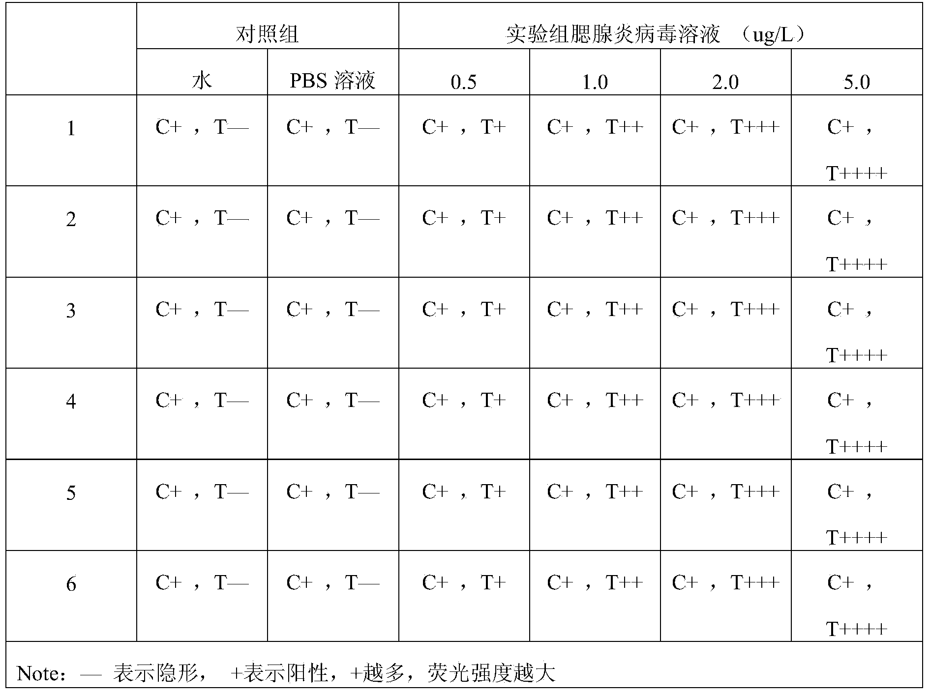
[0058] Embodiment 3: detect mumps virus with described test paper, comprise the following steps: spot sample on the assembled test paper close to one end of mumps virus IgG monoclonal antibody, after reaction 5min, observe result in ultraviolet analyzer . PBS buffer solution and normal human blood were used as blank controls.
[0059] Result judgment: under the premise that the C band shows a red fluorescent band, the intensity of the fluorescent band of the T band is visually compared with the blank. The weaker the fluorescence, the lower the concentration of the tested substance in the test solution.
PUM
 Abstract
Description
Claims
Application Information
 - R&D Engineer
- R&D Manager
- IP Professional
- Industry Leading Data Capabilities
- Powerful AI technology
- Patent DNA Extraction
Browse by: Latest US Patents, China's latest patents, Technical Efficacy Thesaurus, Application Domain, Technology Topic, Popular Technical Reports.
© 2024 PatSnap. All rights reserved.Legal|Privacy policy|Modern Slavery Act Transparency Statement|Sitemap|About US| Contact US: help@patsnap.com