Microfluidic chip apparatus and application thereof
A microfluidic chip and chamber technology, applied in the direction of microorganisms, laboratory containers, microorganism measurement/inspection, etc., to achieve the effect of simple production, good biocompatibility and easy operation
- Summary
- Abstract
- Description
- Claims
- Application Information
AI Technical Summary
Problems solved by technology
Method used
Image
Examples
Embodiment 1
[0041] Embodiment 1: the structure of the chip
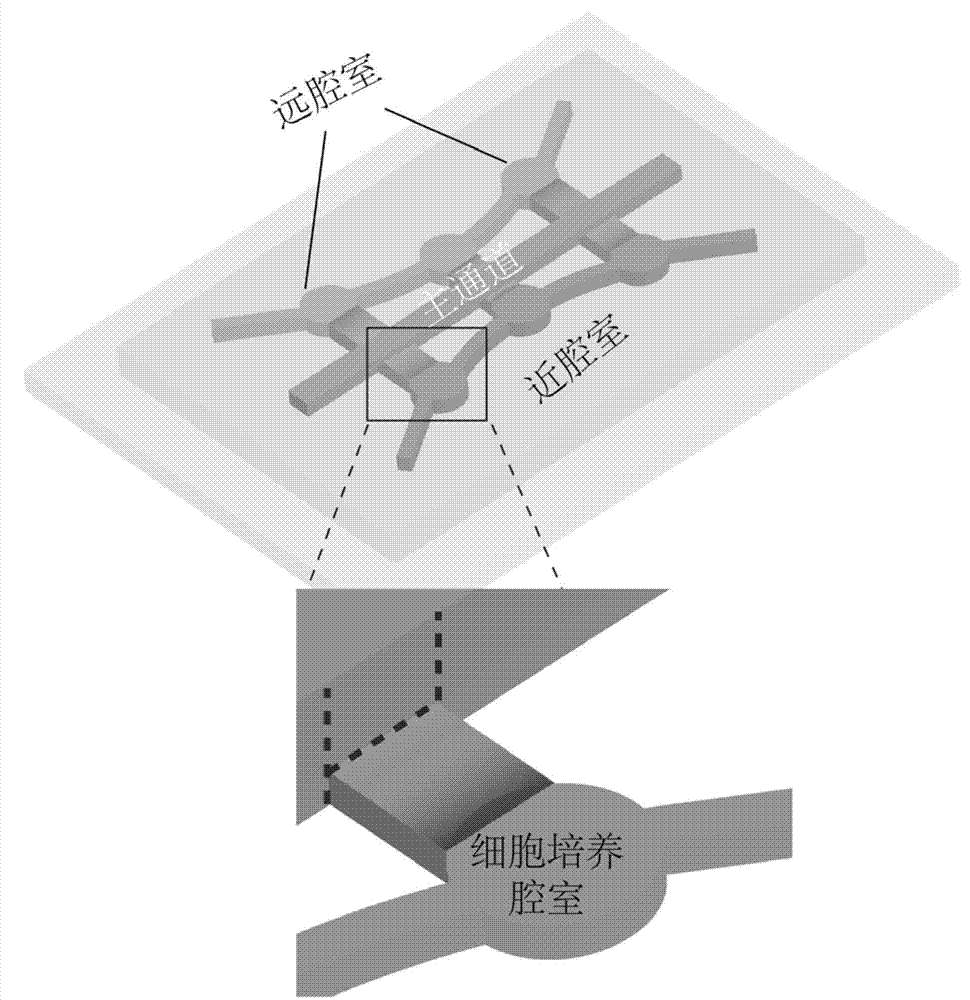
[0042] Such as figure 1 As shown, the microfluidic chip device of the present invention is composed of a main channel and a cell culture chamber. Among them, the main channel can simulate blood vessels for transporting cell culture medium and quantum dot solution, and the cell culture chamber can simulate the adjacent tissue around blood vessels where quantum dots act.
[0043] The device includes two parts: a main channel and a cell culture chamber, and the main channel communicates with the cell culture chamber. The heights of the main channel and the cell culture chamber of the microfluidic chip device are different, 71 μm and 38 μm, respectively. In this way, the surface tension effect can be used to prevent the mixture of cells and three-dimensional culture substrate from leaking into the main channel when injected into the cell culture chamber. The cell culture chamber of the microfluidic chip device is divided into two...
Embodiment 2
[0044] Example 2: Modeling Diffusion in Tissue
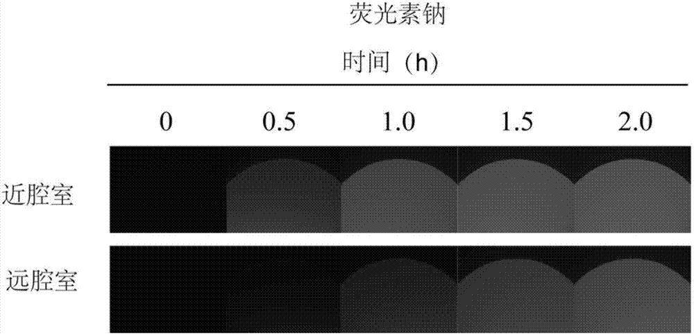
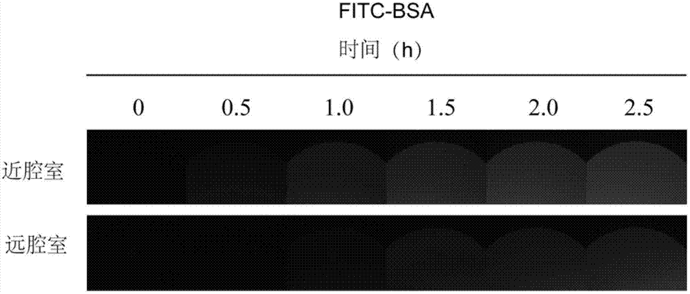
[0045] Diffusion in tissues using sodium fluorescein and fluorescein isothiocyanate-labeled bovine serum albumin (FITC-BSA) as model drugs. Inject agarose solution with a mass volume ratio of 0.5% into the cell culture chambers on both sides. After it coagulates, inject 100 μM sodium fluorescein solution and 2.75 mg / mL FITC-BSA solution from the main channel respectively. At the same position on the top of the chamber, a fluorescent microscope (Leica DMI 4000B) was used to take pictures of the fluorescence intensity of sodium fluorescein every 5 minutes, and the fluorescence intensity of FITC-BSA every 10 minutes. The experiment of fluorescein sodium lasted for 120 minutes, and the experiment of FITC-BSA lasted for 300 minutes. The results are as follows figure 2 and image 3 shown. From Figure 2-4 It can be seen that the fluorescence intensity becomes stronger with time, indicating that the two substances have indeed diff...
Embodiment 3
[0046] Example 3: Three-dimensional culture of HepG2 cells in a chip
[0047] Two plates 60cm 2 The overgrown cells in the culture dish were digested, the cell suspension was centrifuged, and the supernatant was removed, and the remaining cells were redispersed to a cell density of 10 6 / mL. The three-dimensional culture matrix was prepared by mixing 100 μL 3% (w / v) low melting point agarose solution, 100 μL fetal bovine serum and 100 μL phosphate buffer solution. Equal volumes of cell suspension and three-dimensional culture matrix are mixed and injected into the cell culture chamber of the microfluidic chip of the present invention, and the chip is placed at 4°C for 10 minutes to accelerate the solidification of the agarose and wrap the cells in it. Finally, inject the culture medium into the main channel and coat the surface of the chip to prevent the volatilization of the culture medium in the channel. The chip was placed in a cell incubator, the medium was changed ever...
PUM
 Abstract
Description
Claims
Application Information
 - R&D Engineer
- R&D Manager
- IP Professional
- Industry Leading Data Capabilities
- Powerful AI technology
- Patent DNA Extraction
Browse by: Latest US Patents, China's latest patents, Technical Efficacy Thesaurus, Application Domain, Technology Topic, Popular Technical Reports.
© 2024 PatSnap. All rights reserved.Legal|Privacy policy|Modern Slavery Act Transparency Statement|Sitemap|About US| Contact US: help@patsnap.com


