Method for separating and purifying mesenchymal stem cells originated from formation tissue
A mesenchymal stem cell and tissue technology, applied in the field of separation and purification of mesenchymal stem cells, can solve the problems of cumbersome separation and purification methods, high cost, and inapplicability of mesenchymal stem cell separation and purification, achieve good sorting effect and improve purification efficiency Effect
- Summary
- Abstract
- Description
- Claims
- Application Information
AI Technical Summary
Problems solved by technology
Method used
Image
Examples
Embodiment 1
[0037] Take the decidua tissue on the maternal side of the placenta, cut it into small pieces, take 5g of the fragments and add it to 10mL containing 0.1% (w / v, ie 1g / L, 1g of enzyme per 1L of digestive juice) type IV collagenase (Sigma company, 125U / mg) in the digestive solution (distilled solution solvent is double-distilled water) and digested for 30 minutes to collect the cells, with cell culture medium (adding 10% fetal bovine serum (Hangzhou Sijiqing Bioengineering Materials Co., Ltd.) and 2mM L-glucose Aminoamide (Sigma Company) was suspended in DMEM-LG (Hyclone Company) culture medium (prepared by adding 100 mL fetal bovine serum and 2 mmol L-glutamine to 900 mL of basic DMEM-LG culture liquid, the same below), and the suspended cells were collected for different Time period of adherent culture.
[0038]1. After the collected suspension cells are cultured in a 6-well culture plate for 8 hours, collect the unattached suspension cells, keep the cells that have been attac...
Embodiment 2
[0044] Example 2: Screening of clones of cells:
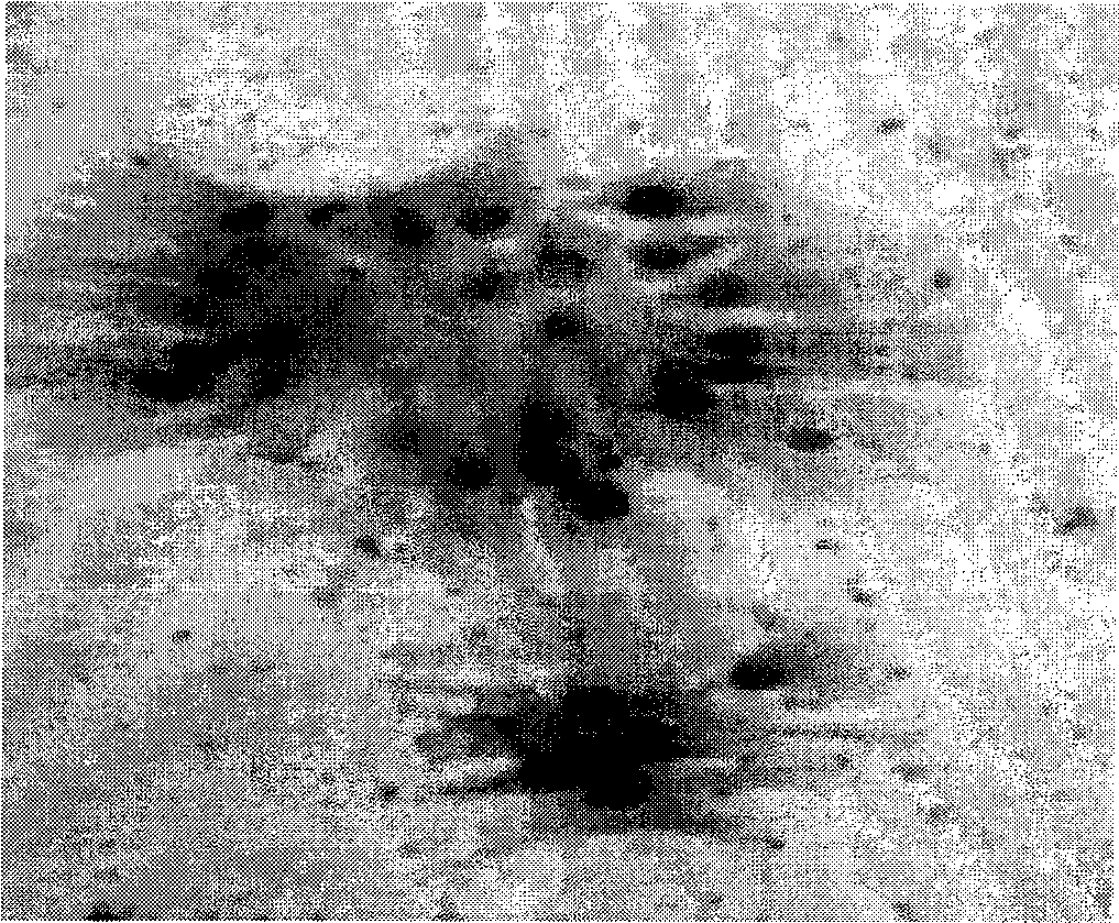
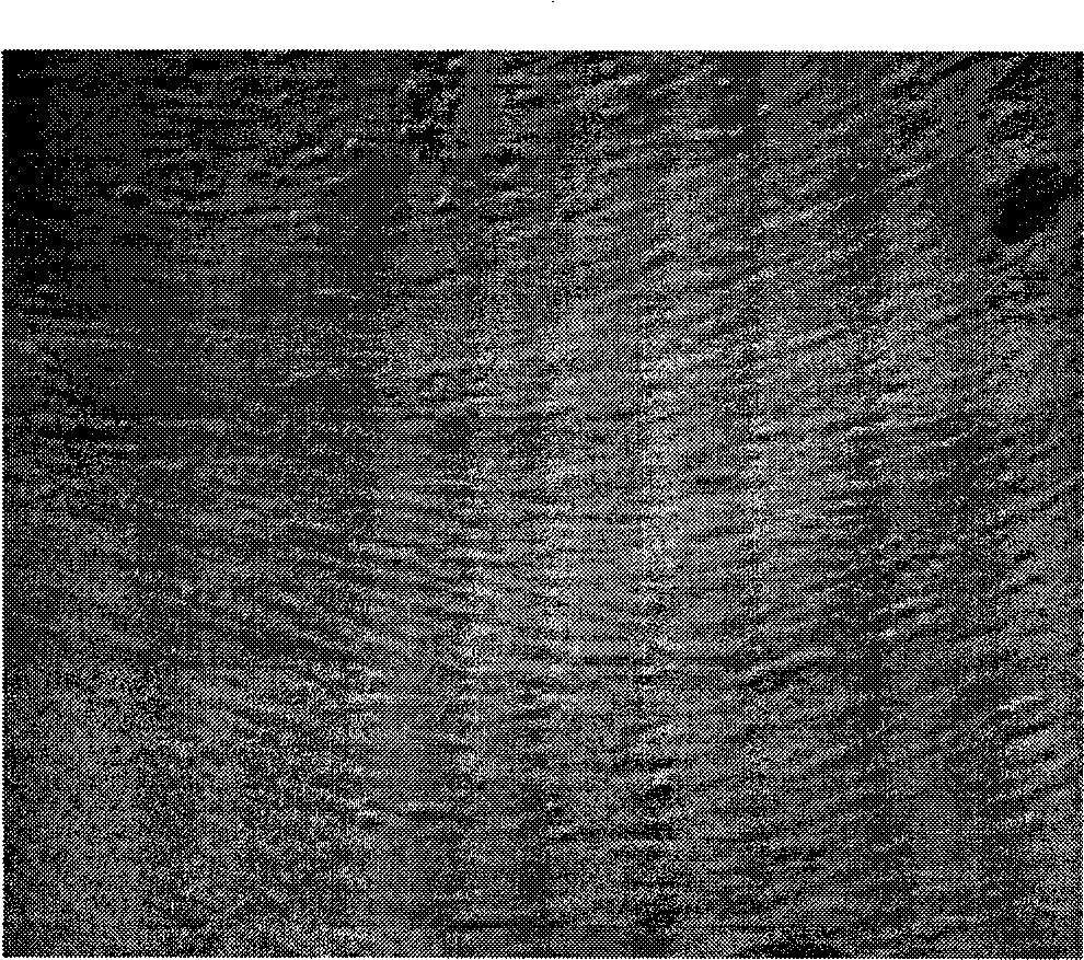
[0045] According to the results of Example 1, the cells sorted by the optimal adherence time period (72 hours) were screened for cell clone culture. Attached cells were digested with Tryspin-EDTA and harvested for a period of 72 hours. The harvested cells were washed once with PBS and then suspended in the cell culture medium (preparation: 100mL fetal bovine serum, 2mmol L-glutamine, 900mL basic DMEM-LG medium mixed), and carried out cell density gradient release by limiting dilution method Afterwards, cell clones were cultured in 96-well culture plates. After the cell clones are formed, the clones of single cells are collected, and then transferred to 24-well culture plates for subculture and expansion culture. See Figure 1(A) for the cell clone formed by a single cell; Figure 1(B) for the cell morphology after the cloned cells are expanded and layered in a 24-well culture plate, which is long fiber-like cells.
[0046] The...
Embodiment 3
[0047] Embodiment 3: comparative analysis of amplification ability
[0048] Collect the cells attached to the best adherent time period (72 hours), and placenta tissue cells digested with 0.1% type IV collagenase directly adhere to the adherent cells in a 6-well culture plate and culture them for 72 hours (not carried out). Adhered cells sorted by time-period adherence) were used as control, cultured in a 24-well culture plate with cell culture medium (preparation: 100mL fetal bovine serum, 2mmol L-glutamine, 900mL basal DMEM-LG medium mixed) , and assayed for cell expansion capacity. Experimental results such as figure 2 , where —◇—indicates adherent cells that have not been sorted by time-period adherence, and —◆—indicates cells that adhere to the best adherence time period (72 hours).
[0049] From figure 2 According to the analysis of the comparison results, the cells attached in the best time period (72 hours) have a significant growth advantage: the cells grow rapidly...
PUM
 Abstract
Description
Claims
Application Information
 - R&D
- Intellectual Property
- Life Sciences
- Materials
- Tech Scout
- Unparalleled Data Quality
- Higher Quality Content
- 60% Fewer Hallucinations
Browse by: Latest US Patents, China's latest patents, Technical Efficacy Thesaurus, Application Domain, Technology Topic, Popular Technical Reports.
© 2025 PatSnap. All rights reserved.Legal|Privacy policy|Modern Slavery Act Transparency Statement|Sitemap|About US| Contact US: help@patsnap.com


