
Method for detecting protein content difference

A technology of protein content and determination method, which is applied in the field of relative protein content, can solve problems such as inconvenient and effective detection methods and cumbersome determination, and achieve the effect of less reagent consumption and low sample consumption

Inactive Publication Date: 2009-03-11
周国华
View PDF0 Cites 14 Cited by
  • Summary
  • Abstract
  • Description
  • Claims
  • Application Information

AI Technical Summary

Problems solved by technology

However, this method relies on real-time fluorescent PCR quantification, requires an expensive real-time fluorescent PCR instrument, and needs to make a standard curve, which is cumbersome to measure. More importantly, only one sample can be measured at a time. Or the difference in expression levels in three different cells needs to be measured three times, and then compared and analyzed. Therefore, it is not a convenient and effective detection method

Method used


Image

  • Method for detecting protein content difference
  • Method for detecting protein content difference
  • Method for detecting protein content difference

Examples


Embodiment 1

[0039] Embodiment 1: the determination of the relative content of interleukin 2 (IL-2) of two kinds of different sources

[0040] 1. Experimental materials: recombinant human interleukin-2 (Peprotech, USA); biotinylated anti-human interleukin-2 polyclonal antibody (R&D System, USA); maleimide-modified streptavidin (Sigma, USA); Magnetic beads (DynabeadsM-280-strptavidin, Dynal AS, Oslo, Norway); oligonucleotide single-stranded (Invitrogen synthesis, Shanghai) Probe 1: 5'-SH-ttttt ttatg tggtc tatgt cgtcg ttcgc tagta gttcc tgggc tgcac- 3'; Probe 2: 5'-P-tcgaggcgta gaatt ccccc gatgc gcgct gttct tactc atttt t-SH-3'; Sequence tag: 5'-P-agt tgc cat ctg tcgatc-3', 5'-P-agt tgc cat ctg tca ctg-3'; linking single strand: 5'-acgcc tcgac agtga cagat ggcaa ctgtgcagcc cttt-3', 5'-acgcc tcgag atcga cagat ggcaa ctgtg cagcc cttt-3'; amplification primer: 5'-Bio -atgtggtcta tgtcg tcgtt cg-3', 5'-tgagt aagaa cagcg cgcat-3'; annealing primer: 5'-ggggg aattc tacgcctcga-3'.

[0041] 2. Experimen...

Embodiment 2

[0052] Example 2: Determination of protein content differences by directly labeling source-specific sequences on DNA molecules linked to antibody proteins

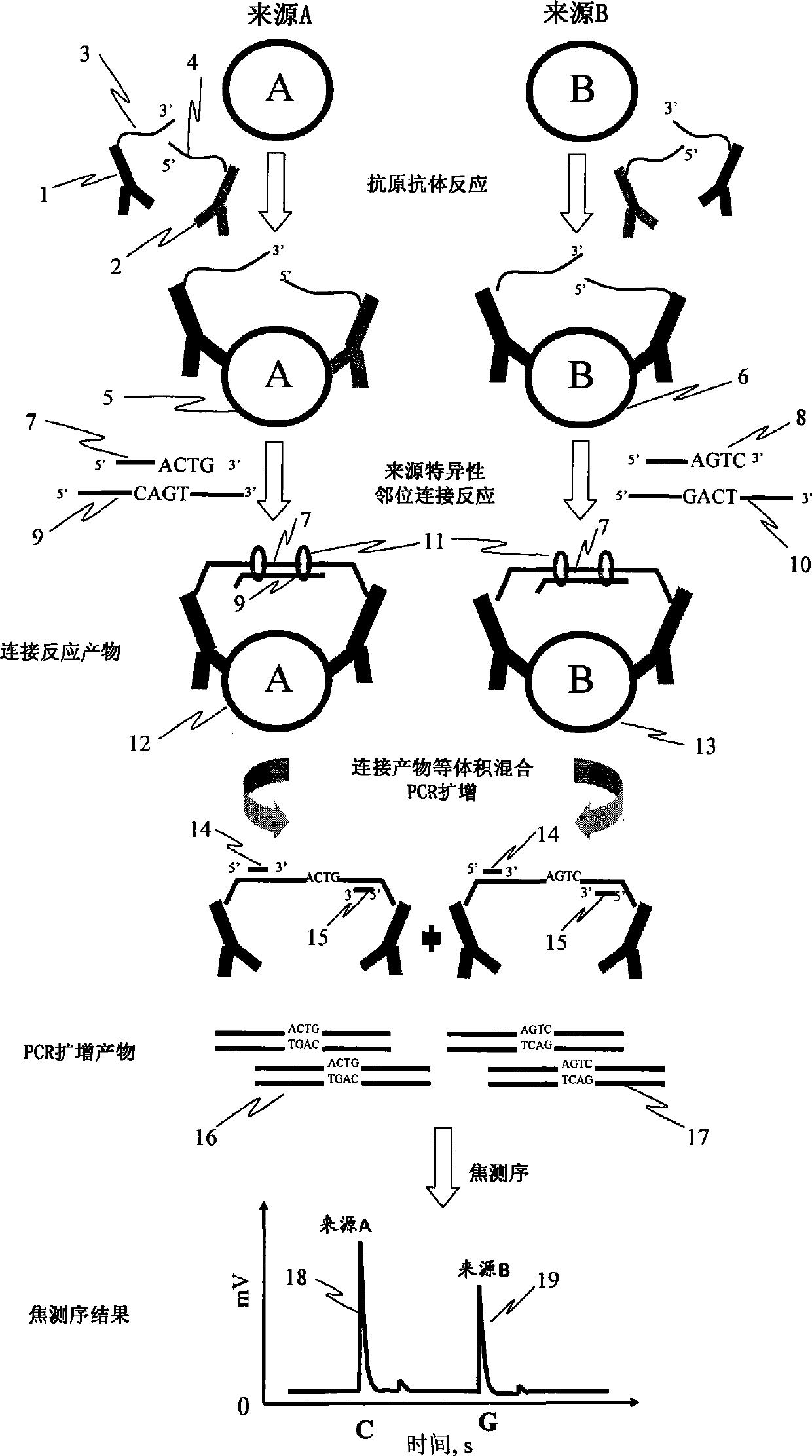
[0053] In this example, the figure 1 The source-specific base sequences in DNA single strands 7 and 8 are marked in DNA sequence 3 or 4 respectively, that is, the DNA sequences in antigen-antibody-DNA complexes 5 and 6 formed after the antigen-antibody reaction are different and contain source-specific sexual sequence. When performing the source-specific proximity ligation reaction, it is only necessary to add a single-stranded DNA 9 complementary to the sequences in 3 and 4 for the ligation reaction. The rest of the steps are the same as figure 1 unanimous.

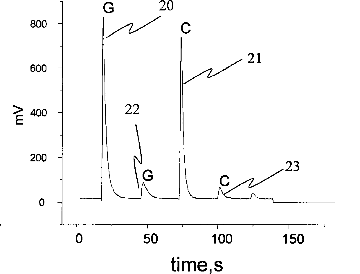
[0054] In this example, DNA with different sequences needs to be labeled with the same antibody to distinguish proteins from different sources. The determination procedure is the same as that in Example 1. Since the content of IL2 from the two sources is equal, t...

Embodiment 3

[0055] Example 3: The method of adding a third antibody to reduce the background signal of the reaction and improve the sensitivity of the assay

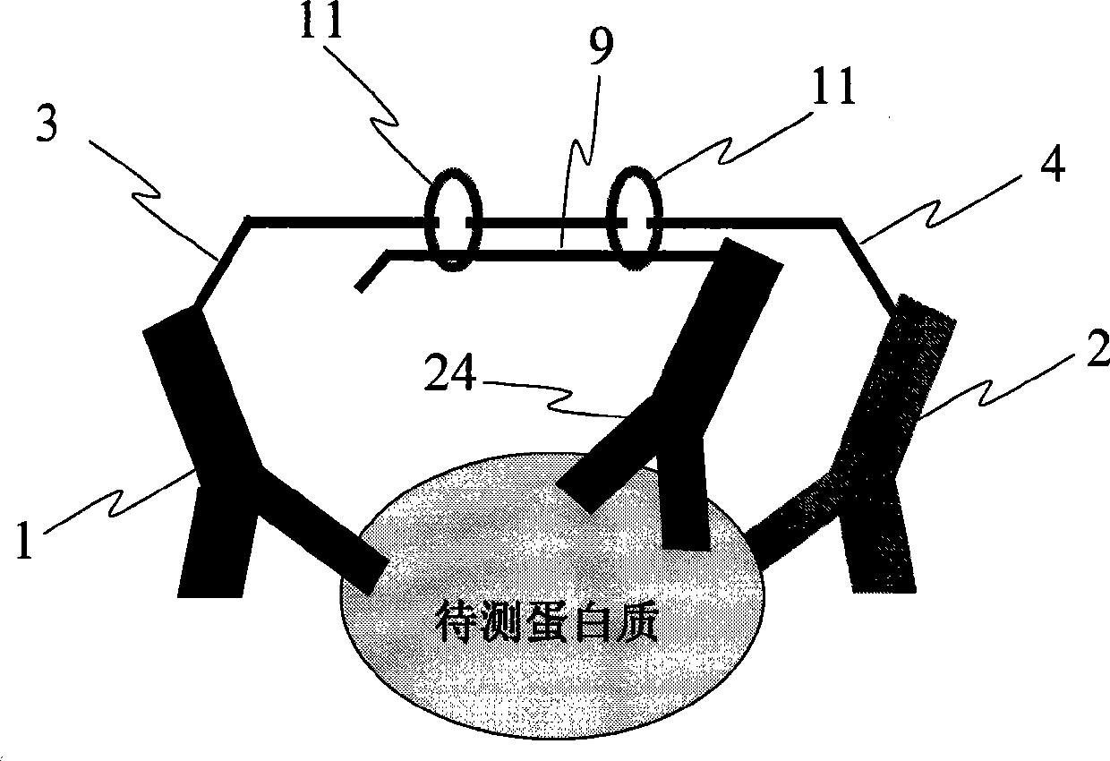
[0056] In this embodiment, at least three antibody-DNA composite probes are prepared, and the antibodies are antibodies to the same protein. Such as image 3 shown, except figure 1 In addition to the antibodies 1 and 2, an antibody 24 should be added, and the adjacent linked DNA single strand (9) containing the source-specific sequence should be connected with the antibody 24. After the antigen-antibody reaction occurs, the ligation reaction occurs under the action of ligase 11. This results in increased specificity and reduced background in the ligation reaction.


PUM


Abstract

The invention discloses a method for determining the differences of protein contents, which comprises the steps that: a. a plurality of kinds of protein antibodies respectively combined with different oligonucleotide sequences synchronously carry out antigen antibody reaction with one kind of proteantigen to be tested; b. the oligonucleotide sequences of the antigen antibody complex in the step a are coupled together through the coupled reaction by using ligase, and the sequences of a ligation product are led to contain a base sequence representing the source of the protein to be tested; c. the ligation product in the step b is taken as a templet, and a base sequence containing the specificity of the source of the protein to be tested is amplified by adopting the amplification reaction; d. the base sequence representing the source of the protein to be tested in the amplification product sequence is determined by adopting the real-time sequencing technology; and e. the sequence determined in the step d is taken as the source of the protein to be tested, signal intensity of each sequence is compared to obtain the relative contents of the protein to be tested in each source. The method can determine the differences of protein expression content from different sources at tone time with low cost.

Description

technical field [0001] A method for determining the difference in protein content of the present invention belongs to a method for determining relative protein content, specifically a method for detecting protein content through nucleic acid-labeled protein reaction, antigen-antibody reaction, enzyme ligation reaction, nucleic acid amplification reaction and real-time sequencing reaction To determine the relative protein content of different sources. Background technique [0002] The Human Genome Project was completed ahead of schedule due to the development of capillary array electrophoresis sequencing technology (Anal Chem 74(1):22A, 2002). Like capillary array electrophoresis, the development of two-dimensional electrophoresis technology, especially chromatography-mass spectrometry technology, has also greatly accelerated the pace of proteomics research (Lambert, J.P., M.Ethier, et al.Anal Chem 77(12): 3771-87, 2005). However, protein molecules are much more complex tha...

Claims


Application Information

Patent Timeline
Patent Type & Authority Applications(China)
IPC IPC(8): G01N33/68G01N33/577
Inventor 周国华
Owner 周国华
Learn More