
Antibodies against kidney associated antigen 1 and antigen binding fragments thereof

an antigen and antigen technology, applied in the field of antibodies or antigen binding fragments, can solve the problems of high mortality rate, limited clinical application of early stage disease tests, and high cost and availability of strategies, so as to improve binding and internalization, improve the ability to bind, and reduce the affinity

Active Publication Date: 2015-01-20
ADC THERAPEUTICS SA
View PDF231 Cites 6 Cited by
  • Summary
  • Abstract
  • Description
  • Claims
  • Application Information

AI Technical Summary

Benefits of technology

These antibodies demonstrate enhanced binding affinity and internalization capabilities, potentially leading to improved therapeutic outcomes with reduced side effects and improved detection accuracy for ovarian cancer and other cancer types.

Problems solved by technology

The high mortality rate is due to the difficulties with the early detection of ovarian cancer when the malignancy has already spread beyond the ovary.
However, this strategy is expensive and not broadly available.
Consequently, this test has very limited clinical application for the detection of early stage disease when it is still treatable, exhibiting a positive predictive value (PPV) of <10%.
Thus, this test is not an effective screening test.
Poor outcomes have been attributed to (1) lack of adequate screening tests for early disease detection in combination with only subtle presentation of symptoms at this stage—diagnosis is frequently being made only after progression to later stages, at which point the peritoneal dissemination of the cancer limits effective treatment and (2) the frequent development of resistance to standard chemotherapeutic strategies limiting improvement in the 5-year survival rate of patients.
However, severe side effects often lead to an incomplete course of treatment.
Notwithstanding these recent advances in the understanding and the treatment for ovarian cancer, the use of chemotherapy is invariably associated with severe adverse reactions, which limit their use.

Method used


Image

  • Antibodies against kidney associated antigen 1 and antigen binding fragments thereof
  • Antibodies against kidney associated antigen 1 and antigen binding fragments thereof
  • Antibodies against kidney associated antigen 1 and antigen binding fragments thereof

Examples


example 1

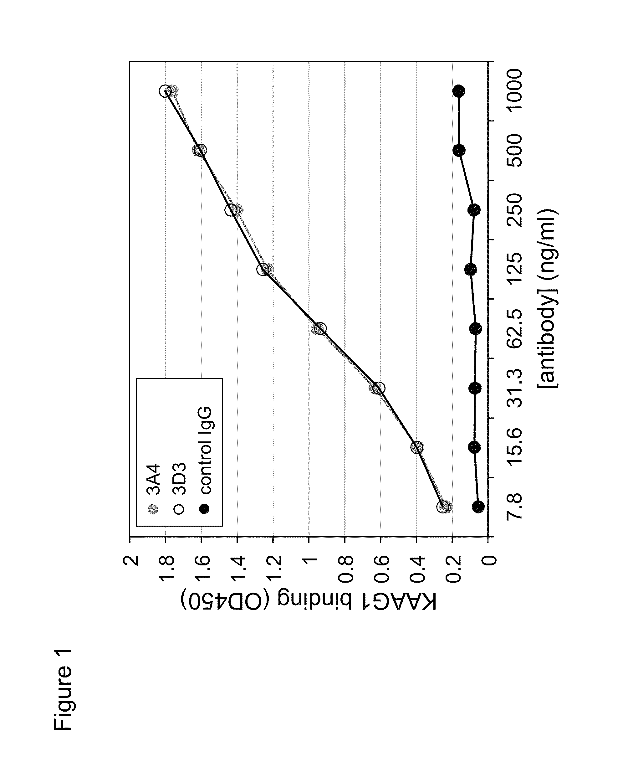
This Example Describes the Binding of Antibody 3A4 to KAAG1

[0323]The antibodies that bind KAAG1 were generated using the Alere phage display technology. A detailed description of the technology and the methods for generating these antibodies can be found in the U.S. Pat. No. 6,057,098. In addition, a detailed description of the generation of antibodies against KAAG1 can be found in PCT / CA2009 / 001586. Briefly, the technology utilizes stringent panning of phage libraries that display the antigen binding fragments (Fabs). After a several rounds of panning, a library, termed the Omniclonal, was obtained that was enriched for recombinant Fabs containing light and heavy chain variable regions that bound to KAAG1 with very high affinity and specificity. From this library, more precisely designated Omniclonal AL0003 A2ZB, 96 individual recombinant monoclonal Fabs were prepared from E. coli and tested for KAAG1 binding. The monoclonal designated 3A4 was derived from this 96-well plate of mon...

example 2

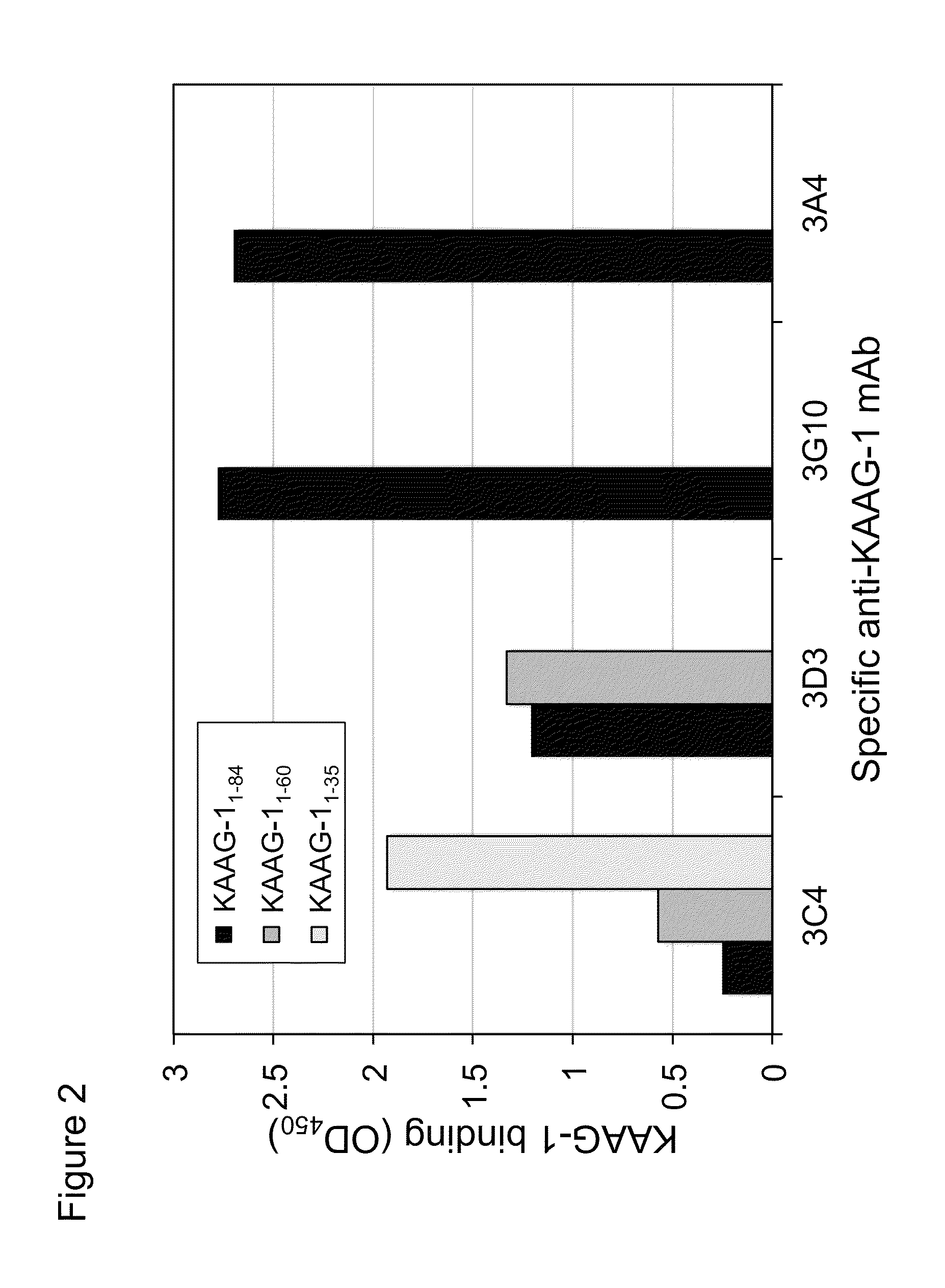
This Example Describes the Epitope Mapping Studies to Determine which Region of KAAG1 the 3A4 Antibody Binds to

[0329]To further delineate the regions of KAAG1 that are bound by the 3A4 antibody, truncated mutants of KAAG1 were expressed and used in the ELISA. As for the full length KAAG1, the truncated versions were amplified by PCR and ligated into BamHI / HindIII digested pYD5. The primers that were used combined the forward oligonucleotide with the sequence shown in SEQ ID NO.:25 with primers of SEQ ID NOS:26 and 27, to produce Fc-fused fragments that ended at amino acid number 60 and 35 of KAAG1, respectively. The expression of these recombinant mutants was conducted as was described above for the full length Fc-KAAG1 and purified with Protein-A agarose.

[0330]Based on the teachings of Tremblay and Filion (2009), it was known that other antibodies interacted with specific regions of recombinant KAAG1. Thus, anti-KAAG1 antibody 3C4, 3D3, and 3G10 interacted with the regions 1-35, 36...

example 3

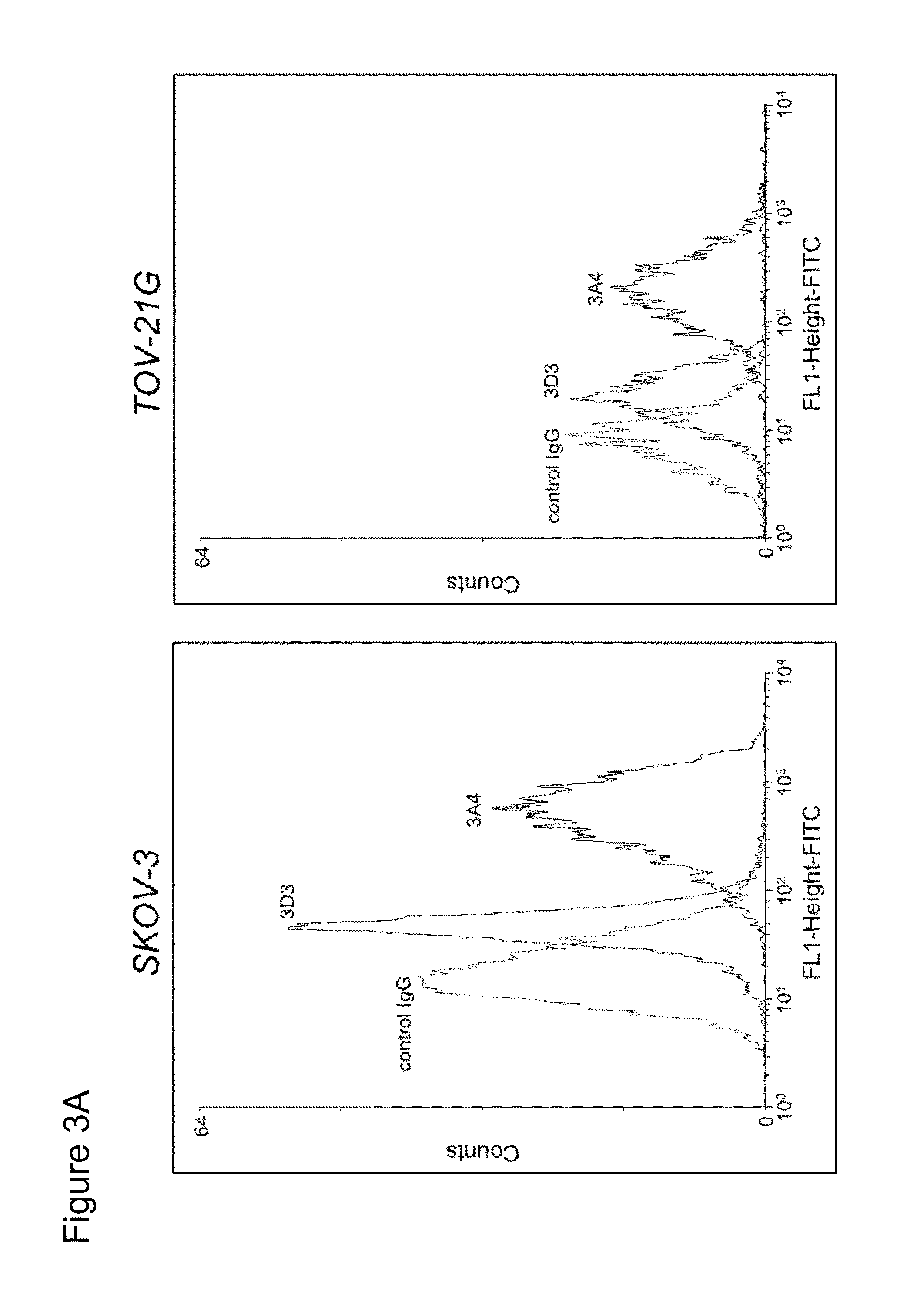
This Example Describes the Ability of 3A4 to Bind to KAAG1 on the Surface of Cancer Cell Lines

[0331]Flow cytometry was used to detect KAAG1 on the surface of cell lines. Based on RT-PCR expression analyses using KAAG1 mRNA specific primers, selected cancer cell lines were expected to express KAAG1 protein. To verify this, ovarian cancer cells (SKOV-3 and TOV-21G) and a control cell lines that showed very little KAAG1 expression (293E). The cells were harvested using 5 mM EDTA, counted with a hemocytometer, and resuspended in FCM buffer (0.5% BSA, 10 μg / ml goat serum in 1×PBS) at a cell density of 2×10⁶ cells / ml. Chimeric 3A4 or a control IgG were added to 100 μl of cells at a final concentration of 5 μg / ml and incubated on ice for 2 h. The cells were washed in cold PBS to remove unbound antibodies, resuspended in 100 μl FCM buffer containing anti-human IgG conjugated to FITC (diluted 1:200) as a secondary antibody and incubated on ice for 45 min on ice. Following another washing ste...


PUM


Abstract

Novel antibodies and antigen binding fragments that specifically bind to KAAG1 and which may be used in the treatment, detection and diagnosis of cancer comprising KAAG1-expressing cells are disclosed herein. Cells expressing the antibodies and antigen binding fragments as well as methods of detecting and treating cancer using the antibodies and fragments are also disclosed. Cancer indications which may benefit from such treatment or detection include ovarian cancer, renal cancer, lung cancer, colorectal cancer, breast cancer, brain cancer, and prostate cancer, as well as melanomas.

Description

PRIORITY CLAIM[0001]This patent application is a national stage filing under 35 U.S.C. §371 of international application No. PCT / CA2012 / 000296 filed on Mar. 28, 2012 which claimed priority to U.S. provisional application No. 61 / 470,063 filed Mar. 31, 2011 and U.S. provisional application No. 61 / 533,346 filed on Sep. 12, 2011. The entire contents of each of these priority applications are incorporated herein by reference.SEQUENCE LISTING[0002]In accordance with 37 C.F.R. §1.52(e)(5), a Sequence Listing in the form of a text file (entitled “Sequence Listing”, created on Sep. 25, 2013 of 90 kilobytes) is incorporated herein by reference in its entirety.FIELD OF THE INVENTION[0003]The present invention relates to specific antibodies or antigen binding fragments that specifically bind to kidney associated antigen 1 (KAAG1) and their use for the treatment, detection and diagnosis of cancer. Delivery of a therapeutic agent to cells with these antibodies or antigen binding fragments is part...

Claims


Application Information

Patent Timeline
Patent Type & Authority Patents(United States)
IPC IPC(8): A61K47/48A61K39/395C07K16/00G01N33/574C07K16/30
CPCC07K2317/92C07K2317/55C07K16/00C07K2317/56C07K2317/34C07K2317/567C07K2317/24A61K47/48607C07K16/3069C07K16/3038G01N33/57484A61K47/48638A61K39/39558A61K38/06A61K45/06C07K16/18C07K2317/21C07K2317/565C07K16/30C07K16/3015C07K16/3023C07K16/3053A61K47/6861A61K47/6869A61P35/00A61P35/02A61P35/04C07K16/28C07K2317/515G01N33/57492A61K2039/505C07K2317/74C07K2317/51G01N2333/705
Inventor TREMBLAY, GILLES BERNARDMORAITIS, ANNA N.SULEA, TRAIANFILION, MARIO
Owner ADC THERAPEUTICS SA
Learn More