
Improving fracture toughness of medical devices with a stereocomplex nucleating agent

a nucleating agent and stereocomplex technology, applied in the field of stents, can solve the problems of difficult treatment, adverse or even toxic side effects, and significant problem of stenosis, and achieve the effect of improving the toughness of fractures and improving the toughness of medical devices

Active Publication Date: 2012-12-25
ABBOTT CARDIOVASCULAR
View PDF22 Cites 6 Cited by
  • Summary
  • Abstract
  • Description
  • Claims
  • Application Information

AI Technical Summary

Problems solved by technology

Yet, restenosis remains a significant problem.
When restenosis does occur in the stented segment, its treatment can be challenging, as clinical options are more limited than for those lesions that were treated solely with a balloon.
Effective concentrations at the treated site require systemic drug administration which often produces adverse or even toxic side effects.
Thus, erodible portions of the device can disappear or substantially disappear from the implant region after the treatment regimen is completed.
However, there are potential shortcomings in the use of polymers, such as insufficient fracture toughness, as a material for implantable medical devices, such as stents.

Method used


Image

  • Improving fracture toughness of medical devices with a stereocomplex nucleating agent
  • Improving fracture toughness of medical devices with a stereocomplex nucleating agent
  • Improving fracture toughness of medical devices with a stereocomplex nucleating agent

Examples


example 1

Preparation of PLLA Stent from Extruded and Expanded Tubing Using PLLA / PDLA Stereocomplex as Nucleating Agent

[0096]Step 1 (material mixing): 20 g PDLA (Mw≈600 kg / mol) is mixed with 1 kg of high molecular weight PLLA (Mw≈600 kg / mol) through melt compounding using a twin screw extruder at 200° C., or through solution blending by dissolving both PLLA and PDLA in chloroform and precipitating them into methanol.

[0097]Step 2 (tubing extrusion): The mixed PDLA / PLLA material is extruded in a single screw extruder at 200° C. and directly quenched in cold water. The size of the extruded tubing is set at about 0.02″ for inside diameter (ID) and 0.06″ for outside diameter (OD).

[0098]Step 3 (tubing expansion): The extruded tubing is placed in a glass mold and expanded at about 200° F. to increase its crystallinity and biaxial orientation. The final ID and OD of the expanded tubing are set at 0.10″ and 0.11″, respectively.

[0099]Step 4 (stent preparation): A stent is cut from the expanded tubing u...

example 2

Preparation of PLLA Stent from Extruded Tubing Using PLLA / PDLA Stereocomplex as Nucleating Agent

[0100]Step 1 (material blending): 60 g PDLA (Mw≈600 kg / mol) is blended with 1 kg of high molecular weight PLLA (Mw≈600 kg / mol) through melt compounding in a twin screw extruder at 200° C.

[0101]Step 2 (tubing extrusion): The PDLA / PLLA blend is extruded in a single or twin screw extruder at 200° C. and slowly cooled down in warm / hot water before it's finally quenched in cold water. The size of the extruded tubing is set at about 0.07″ for ID and 0.08″ for OD.

[0102]Step 3 (stent preparation): A stent is directly cut from the extruded tubing using a femto-second laser, crimped down to a smaller size (0.05″) on a balloon catheter and sterilized by an electron beam at a dose of 25 kGray.


PUM

PropertyMeasurementUnit
processing temperatureaaaaaaaaaa
Tgaaaaaaaaaa
Tmaaaaaaaaaa

Abstract

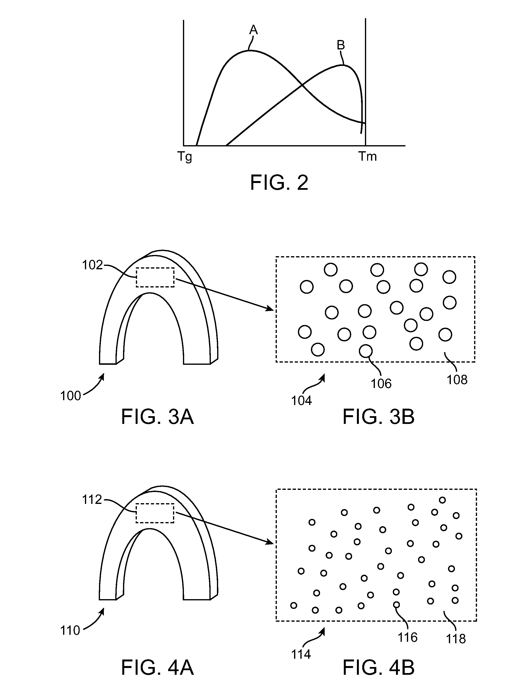
Methods of fabricating a polymeric implantable device from a PLLA / PDLA blend such as a stent with improved fracture toughness are disclosed. The blend is melt processed to allow formation of stereocomplex crystallites, which are nucleation sites for crystal growth. A polymer construct is formed from the melt processed blend and device is formed from the polymer construct. The stereocomplex crystallites result in an in increase in nucleation density and reduced crystal size, which increases fracture toughness of the formed device.

Description

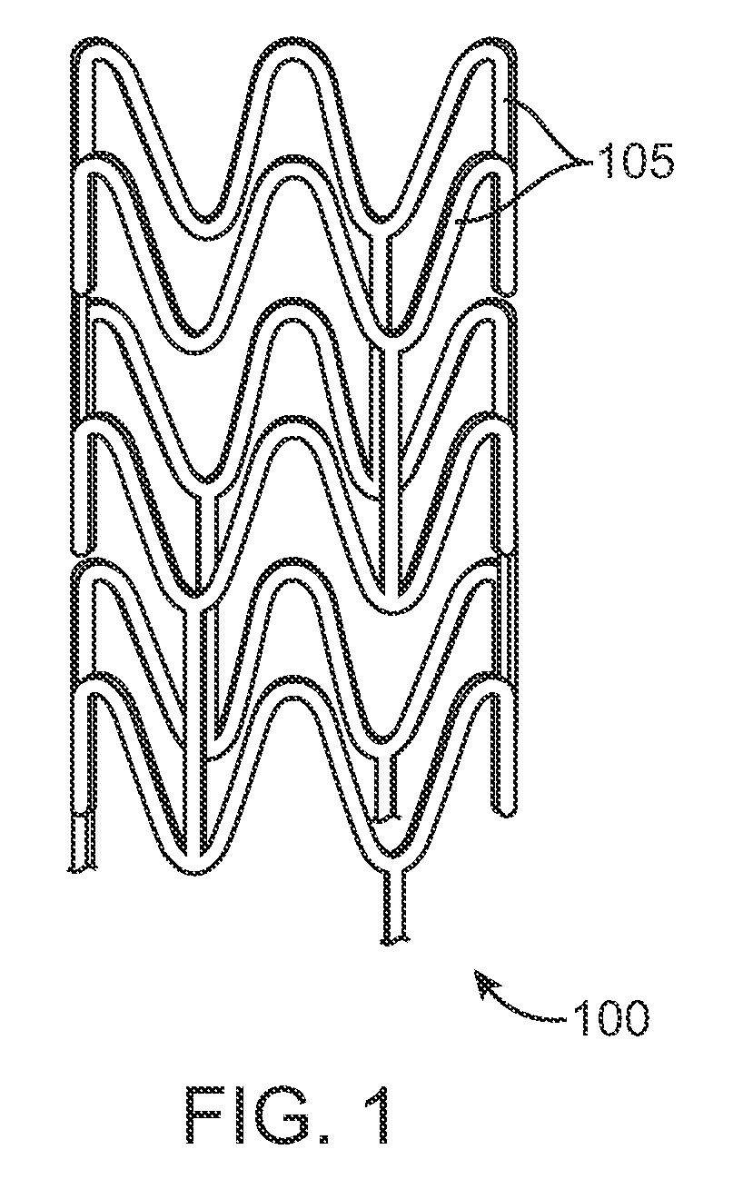
BACKGROUND OF THE INVENTION[0001]1. Field of the Invention[0002]This invention relates to methods of manufacturing polymeric medical devices, in particular, stents.[0003]2. Description of the State of the Art[0004]This invention relates to radially expandable endoprostheses, that are adapted to be implanted in a bodily lumen. An “endoprosthesis” corresponds to an artificial device that is placed inside the body. A “lumen” refers to a cavity of a tubular organ such as a blood vessel. A stent is an example of such an endoprosthesis. Stents are generally cylindrically shaped devices that function to hold open and sometimes expand a segment of a blood vessel or other anatomical lumen such as urinary tracts and bile ducts. Stents are often used in the treatment of atherosclerotic stenosis in blood vessels. “Stenosis” refers to a narrowing or constriction of a bodily passage or orifice. In such treatments, stents reinforce body vessels and prevent restenosis following angioplasty in the v...

Claims


Application Information

Patent Timeline
Patent Type & Authority Patents(United States)
IPC IPC(8): B28B21/56A61F2/915B29C48/32
CPCA61L31/041A61L31/148B29C55/24B29D23/001C08L67/04B29L2031/7534B29C47/0004B29C47/0023B29C47/0026B29C47/882B29K2067/00B29K2067/046B29K2995/006B29L2023/007B29C48/022B29C48/09B29C48/10B29C48/9115B29C48/919
Inventor WANG, YUNBINGOBERHAUSER, JAMES
Owner ABBOTT CARDIOVASCULAR
Learn More