
Microfluidic sensor complex structure

a microfluidic and sensor technology, applied in the field of microfluidic sensor complex structure, can solve the problems of difficult mass production of conventional biosensors in lab-on-chips, complicated structure of conventional biosensors, and difficult production of conventional structures at low cost and high reliability, and achieves convenient handling and shows sensitivity and selectivity

Active Publication Date: 2012-12-25
PRECISIONBIOSENSOR INC
View PDF7 Cites 3 Cited by
  • Summary
  • Abstract
  • Description
  • Claims
  • Application Information

AI Technical Summary

Benefits of technology

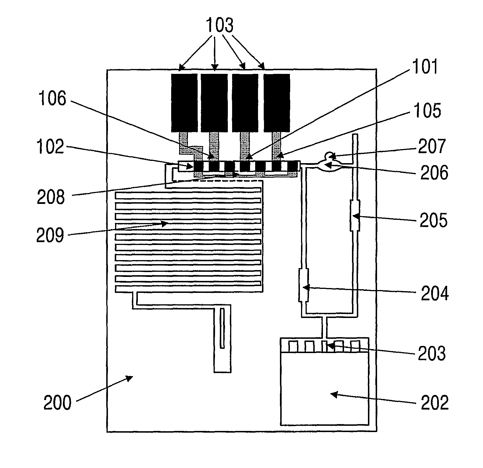
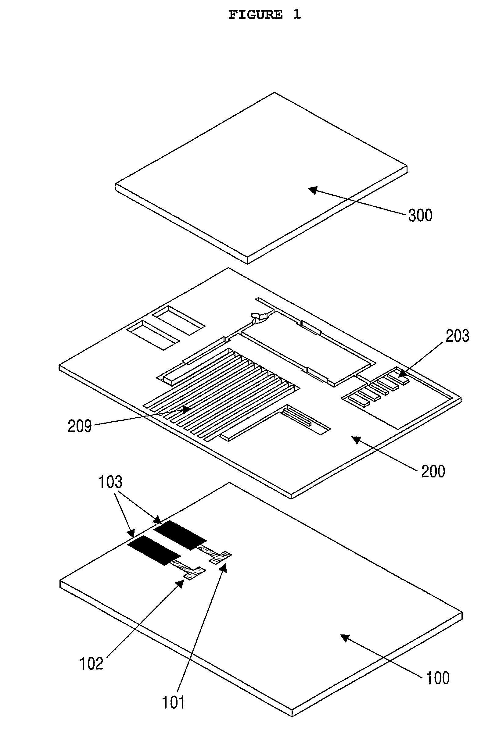
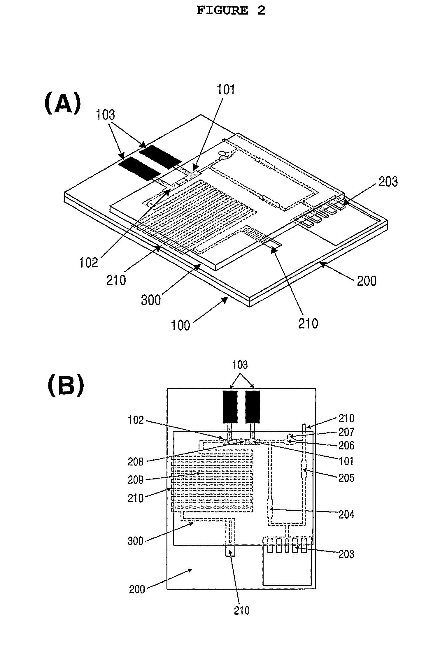
The present invention provides a microfluidic sensor complex structure that is simple, convenient to carry, and can be produced on a mass scale. The structure includes a lower plate with reference electrodes, working electrodes, and fluid sensing electrodes, a middle plate with a sample inlet channel, a microfluidic channel passage, and an upper plate with an air discharge channel and an absorbing channel. The structure also includes a mixing channel and an air inlet channel. The lower plate may also have a fluidity sensing electrode for detecting the arrival of the sample. The middle plate may have a filter pad channel and a filter pad for selectively measuring the analyte component of the sample. The sample flows through the enzyme conjugate reservoir and the substrate reservoir before reaching the detection channel. The detection channel is designed to send quantitative information on the analyte. The absorbing channel maintains a capillary phenomenon to drive the reactions in the detection channel and increase the sensitivity of the detection signals. The method using the microfluidic sensor complex structure allows for quantitative analysis of the analyte.

Problems solved by technology

Conventional lab-on-a-chip sensors, however, have problematic structures in terms of mass production.
In consideration of the fact that lab-on-a-chip technology is applied, in the most part, to disposable biochemical sensors, conventional structures are difficult to produce at low cost and high reliability.
Further, conventional biosensors in lab-on-a-chip are complicated in structure because they use micro-valves, high voltage capillary electrophoresis, and / or combination of complicate miniaturized mechanical components for the delivery of fluid through channels and require washing processes.

Method used


Image

  • Microfluidic sensor complex structure
  • Microfluidic sensor complex structure
  • Microfluidic sensor complex structure

Examples


example 1

Fabrication of Microfluidic Sensor for Quantitative Analysis of Myoglobin 1

[0088](1) Preparation of Electrode and Microfluidic Channel Passage

[0089]A microfluidic channel passage was formed using photolithography, which is generally used in semiconductor technology. In this regard, an acryl-coated polyethylene terephthalate (PET) film was used as a plate. A carbon paste was screen-printed on the film to form a working electrode, an electrode connection and a fluidity sensing electrode. A reference electrode and an electrode connection were formed with a silver paste. A dry photoresist film was thermally pressurized against the film on which the electrodes were formed, followed by the formation of a microfluidic channel passage using an exposure system. The microfluidic channel passage was designed with a CAD program and printed on an OHP film, which was then used as a pattern mask. After being exposed to a UV beam at a predetermined dose, the dry photoresist film was etched with a 2...

example 2

Fabrication of Microfluidic Sensor for Quantitative Analysis of Myoglobin 2

[0093]A microfluidic sensor was fabricated in the same manner as set forth in Example 1, with the exception that a blood filter pad was provided at the sample inlet channel and a fluidity sensing electrode was installed at an end region of the absorbing channel.

[0094]The blood filter pad was commercially available from Lydall Filtration, identified as LyPore Grade 9389. Like the working electrode, the fluidity sensing electrode was prepared from a carbon paste.

example 3

Fabrication of Microfluidic Sensor for Quantitative Analysis of Myoglobin 3

[0095]A microfluidic sensor was fabricated in the same manner as explained in Example 1, with the exception that two verifying electrodes were further installed. The two verifying electrodes were formed of carbon paste and coated with BSA for a first verifying electrode and with IgG for a second verifying electrode.

[0096]Coated with BAS, the first verifying electrode was adapted to detect background signals while the second verifying electrode, coated with the same IgG as in the control line of a rapid kit, was adapted to monitor the solution of the enzyme conjugate by binding to unreacted enzyme conjugate, that is, to monitor saturation signals.


PUM

PropertyMeasurementUnit
temperatureaaaaaaaaaa
pHaaaaaaaaaa
pHaaaaaaaaaa

Abstract

Disclosed is a microfluidic sensor complex structure comprising a lower plate, a middle plate and an upper plate. A reference electrode, a working electrode and an electrode connection are formed on the lower plate. The middle plate comprises a microfluidic channel passage therein. The upper plate is overlaid on the middle plate so as to induce a capillary phenomenon on the microfluidic channel passage formed on in the middle plate. The microfluidic sensor complex structure allows the motion of a sample to be driven only by a capillary phenomenon, without additional operation, and allows an immune response, washing, and electrochemical analysis in one round once a sample is introduced thereinto. Hence, it requires only a short time period for measurement, is convenient to handle, and shows sensitivity and selectivity. Also, it can be produced on a mass scale because it can be formed of typical organic polymers using a simple method. Based on analytical electrochemistry, the microfluidic sensor complex structure can be used as a small-size sensor that can be applied to practice sites.

Description

RELATED APPLICATIONS[0001]This application is a 35 U.S.C. §371 national stage filing of PCT Application No. PCT / KR2008 / 004336 filed on Jul. 24, 2008, which claims priority to, and the benefit of, Korean Patent Application No. 10-2007-0075254 filed on Jul. 26, 2007. The contents of the aforementioned applications are hereby incorporated by reference.TECHNICAL FIELD[0002]The present invention relates to a microfluidic sensor complex structure in which an immune response, a washing process, substrate introduction and an analytical signal generation can be conducted sequentially once a sample is introduced thereinto.BACKGROUND ART[0003]With the greatly increased concern about the quality of life brought by remarkable advances in science and technology, the significance of disease diagnosis and prophylaxis, quality assurance of foods and full monitoring of environment is ever increasing. As a result, there is a great need for quantitative measurement of organic or inorganic analytes of i...

Claims


Application Information

Patent Timeline
Patent Type & Authority Patents(United States)
IPC IPC(8): B67D1/00
CPCB01L3/5027B01L3/502707B01L2200/0621B01L2200/143B01L2300/0645B01L2300/0681B01L2300/0816B01L2300/0867B01L2300/0887B01L2400/0406G01N33/49G01N33/00G01N33/48G01N35/00
Inventor CHOI, MOON HEEJUNG, SEUNG HYEUNKIM, YOUNG HOONPARK, JOO-HEONNAM, HAKHYUNCHA, GEUN SIG
Owner PRECISIONBIOSENSOR INC
Learn More