Compositions and methods for improved glycoprotein sialylation
a glycoprotein and sialylation technology, applied in the field of sialylation, can solve the problems of inacceptable changes in the bioprocess used, decrease in protein glycosylation, and cellular environmen
- Summary
- Abstract
- Description
- Claims
- Application Information
AI Technical Summary
Benefits of technology
Problems solved by technology
Method used
Image
Examples
example 1
Isolation of Additional Sialidase Genes in CHO Cells
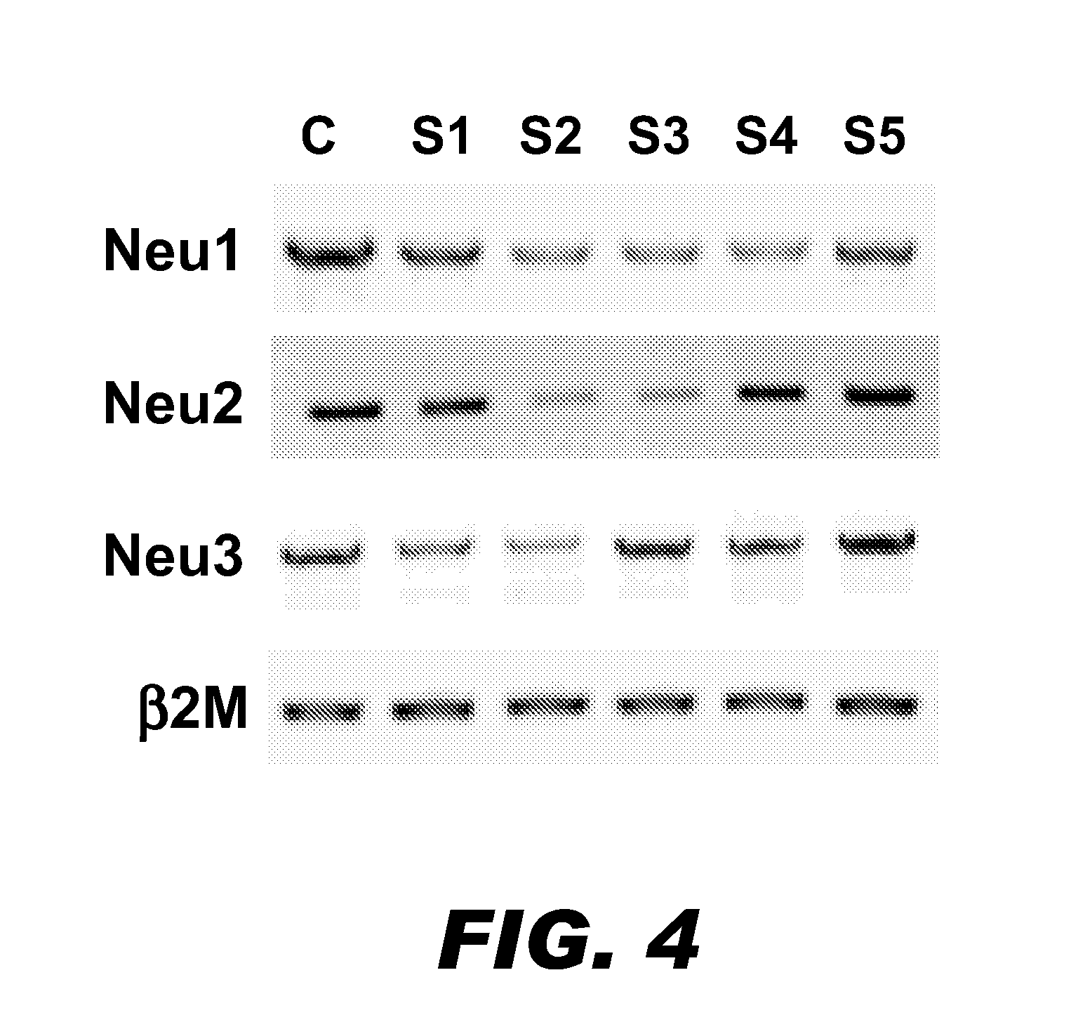
[0074]Four sialidase genes (Neu1, Neu2, Neu3, and Neu4) have been identified and well-characterized in human, mouse, and rat. These sialidases exhibit different enzyme activities, substrate preferences, and subcellular locations. To date, however, only one sialidase gene has been characterized in CHO cells. This gene encodes a cytoplasmic sialidase (i.e., Neu2) (Gramer M J, Goochee C F. (1993) Biotechnol Prog, 9: 366). The purpose of this study was to determine whether CHO cells contain the other sialidase genes.
[0075]Using RT-PCR, 5′ / 3′ RACE (rapid amplification of cDNA ends), and other standard methods, the other three sialidase genes were isolated and cloned from CHO cells. CHO Neu1 encoded a protein with 410 amino acid residues, and CHO Neu3 encoded a protein with 417 amino acid residues (Table 1). On the other hand, CHO Neu4 appeared to be a pseudogene in CHO cells. Both of Neu1 and Neu3 shared high similarities with their cou...
example 2
CHO Neu1 and CHO Neu3 are Sialidases
[0078]Enzymatic sialidase activity studies were performed to confirm that the proteins coded by these two new genes having the expected functions in CHO cells. Briefly, the coding regions for the Neu1 (SEQ ID NO:1) and Neu3 (SEQ ID NO:3) genes, as well as the coding region of the previously reported Neu2 gene (Ferrari et al. (1994) Glycobiology 4(3):367-373) were cloned into pcDNA3.1 mammalian expression vectors. The expression constructs then were transfected into COS7 cells to overexpress the sialidase proteins. The pcDNA3.1 vector alone was also transfected into COS7 cells as the mock transfection. An expression construct containing green fluorescence protein (GFP) was cotransfected into the COS7 cells to normalize the transfection efficiency.
[0079]Twenty-four hours after transfection, the cells were lysed by sonication. The cell debris was removed by centrifugation at 1000×g for 10 min at 4° C. and the supernatant factions were collected for e...
example 3
Subcellular Localization of CHO Sialidases
[0081]To identify the subcellular location for the two new CHO sialidases, immunocytochemistry with Confocal Scanning Microscopy technology was performed. Briefly, the Flag-tag encoding 8 amino acid residues (DYKDDDDK; SEQ ID NO:20) was cloned into the 3′-end of Neu expression constructs to express Flag-tagged fusion proteins. The Flag-tagged constructs were transfected into COS7 cells. Twenty-four hours after transfection, the cells were immunostained using standard procedures.
[0082]The results are presented in FIG. 3. The subcellular locations of the sialidases are indicated by the green fluorescence generated by the FITC-conjugated anti-FLAG antibody. The transfected cells were also immunostained with organelle markers: a lysosomal marker (LysoTracker DND-99), which is indicated by the red fluorescence and a plasma membrane marker (wheat germ agglutinin 647), which is indicated by the blue signal in the FIG. 3. This study revealed that Ne...
PUM
| Property | Measurement | Unit |
|---|---|---|
| nucleic acid sequence | aaaaa | aaaaa |
| solubility | aaaaa | aaaaa |
| thermal stability | aaaaa | aaaaa |
Abstract
Description
Claims
Application Information
 - Generate Ideas
- Intellectual Property
- Life Sciences
- Materials
- Tech Scout
- Unparalleled Data Quality
- Higher Quality Content
- 60% Fewer Hallucinations
Browse by: Latest US Patents, China's latest patents, Technical Efficacy Thesaurus, Application Domain, Technology Topic, Popular Technical Reports.
© 2025 PatSnap. All rights reserved.Legal|Privacy policy|Modern Slavery Act Transparency Statement|Sitemap|About US| Contact US: help@patsnap.com


